¿Cuál es el Dx?

LEARN MSK MRI & SPINE MRI RADIOLOGY ONLINEquiz dx icon circled radedasia


Learn more about various conditions/pathologies in our posts and how to best report it in more detail in our Guided MSK MRI & SPINE MRI Mini-Fellowships.


In our posts we have concise explanation of the MSK MRI & SPINE MRI anatomy, the macroscopic pathology that’s relevant to the Imaging and the Imaging findings in various pathologies.


Learn where to Look, What to Look for and How to Report MRI More Confidently

meniscus cyst radiology intrameniscal tear mri knee

MENISCAL CYST RADIOLOGY INTRAMENISCAL TEAR MRI KNEE

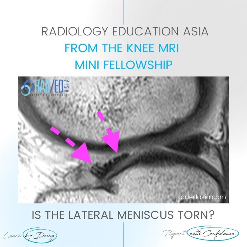
MENISCAL INTRAMENISCAL CYST MRI RADIOLOGY DISCUSSION INTRAMENISCAL CYST. Extensive intra meniscal cyst anterior horn. A tear is not seen extending to the articular margin. However we may not always see the extension on MRI. So if you see an intra-meniscal or parameniscal cyst these are always due to an underlying tear even if we don’t …

MENISCAL CYST RADIOLOGY INTRAMENISCAL TEAR MRI KNEE Leer más »

cord signal change spondylotic myelopathy mri

CORD SIGNAL CHANGE MRI SPONDYLOTIC MYELOPATHY

SPONDYLOTIC MYELOPATHY MRI RADIOLOGY DISCUSSION SPONDYLOTIC MYELOPATHY   Spondylotic Myelopathy is a term used when there is increased cord signal due to extrinsic compression.   Patients will often be symptomatic with cord related symptoms.   By using the term you are indicating that the patient’s symptoms can potentially be reversed by relieving the compression. WHAT …

CORD SIGNAL CHANGE MRI SPONDYLOTIC MYELOPATHY Leer más »

rotator cuff tendinitis mri supraspinatus tendinosis bursitis radiology

ROTATOR CUFF SUPRASPINATUS TENDINOSIS TENDINITIS MRI: WHAT’S WRONG WITH THE SST?

MOVE SLIDER ICON (ARROWS) LEFT TO SEE THE DIAGNOSIS WHAT ARE THE FINDINGS Diffuse Increased signal (intermediate) in the Supraspinatus Tendon. WHAT'S THE Dx SUPRASPINATUS TENDINITIS/TENDINOSIS.  There is diffuse increased signal in the supraspinatus tendon (Pink arrows). The signal increase is intermediate which is seen on tendinitis and not fluid type signal which is seen …

ROTATOR CUFF SUPRASPINATUS TENDINOSIS TENDINITIS MRI: WHAT’S WRONG WITH THE SST? Leer más »

learn knee popliteomeniscal fasicles normal radedasia

POPLITEOMENISCAL FASICLES MRI TEAR NORMAL ANATOMY

POPLITEOMENISCAL FASICLES MRI TEAR NORMAL ANATOMY DISCUSSION These structures are the Popliteomeniscal Fascicles.   They are the Antero Inferior Fascicle (Blue arrow).   The Postero superior Fascicle (Pink arrow) which forms the,   Popliteal Hiatus (Orange arrow) for the,   Popliteus tendon (Green arrow)  to pass through. POPLITEOMENISCAL FASICLES MRI TEAR NORMAL ANATOMY: MOVE SLIDER …

POPLITEOMENISCAL FASICLES MRI TEAR NORMAL ANATOMY Leer más »

patellar tendon trauma contusion mri

PATELLAR TENDON CONTUSION TRAUMA MRI

PATELLAR TENDON CONTUSION Patellar Tendon Contusion. In this case we have Patellar tendon contusion and Patella contusion.   Most commonly this combined appearance would be of Patellar Tendonosis with reactive oedema in the patella and inflammatory change extending into Hoffa’s fat.   However the history of trauma in this patient is important in being able …

PATELLAR TENDON CONTUSION TRAUMA MRI Leer más »

shoulder labrum normal variant buford complex sublabral foramen hole mri

SHOULDER LABRUM NORMAL VARIANTS: WHICH VARIANTS OCCUR HERE (VIDEO)

SHOULDER LABRUM NORMAL VARIANTS DISCUSSION Sublabral Foramen & Buford Complex. FINDINGS Two normal variants occur in superior portion of the anterior labrum: Sublabral Foramen (also called Sublabral Hole), The Buford complex. SHOULDER LABRUM NORMAL VARIANTS VIDEO If your Browser is blocking the video, Please view it on our YouTube Channel HERE. Learn more about SHOULDER …

SHOULDER LABRUM NORMAL VARIANTS: WHICH VARIANTS OCCUR HERE (VIDEO) Leer más »

learn knee mri torn meniscus radedasia

MENISCUS FIBRILLATION MRI LATERAL MENISCUS

MENISCUS FIBRILLATION MRI LATERAL MENISCUS DISCUSSION This is NOT a tear. The anterior horn of the lateral meniscus as it approaches its insertion into the tibia is normally fibrillated like this and this is a normal finding. MENISCUS FIBRILLATION MRI LATERAL MENISCUS: MOVE SLIDER TO VIEW IMAGES ARE FROM OUR INSTAGRAM ACCOUNT. CLICK ICON BELOW …

MENISCUS FIBRILLATION MRI LATERAL MENISCUS Leer más »

Este sitio está destinado únicamente a profesionales médicos. El uso de este sitio se rige por nuestra Términos de servicio y Declaracion de privacidad que se puede encontrar haciendo clic en los enlaces. Acepte antes de continuar con el sitio web.
error: Clic derecho no disponible.