¿Cuál es el Dx?

LEARN MSK MRI & SPINE MRI RADIOLOGY ONLINEquiz dx icon circled radedasia


Learn more about various conditions/pathologies in our posts and how to best report it in more detail in our Guided MSK MRI & SPINE MRI Mini-Fellowships.


In our posts we have concise explanation of the MSK MRI & SPINE MRI anatomy, the macroscopic pathology that’s relevant to the Imaging and the Imaging findings in various pathologies.


Learn where to Look, What to Look for and How to Report MRI More Confidently

freiberg disease mri radiology toe radedasia

FREIBERG DISEASE SUBCHONDRAL FRACTURE INFRACTION OSTEOCHONDROSIS (VIDEO)

FREIBERG DISEASE / SUBCHONDRAL FRACTURE INFRACTION / OSTEOCHONDROSIS DISCUSSION FREIBERG DISEASE / SUBCHONDRAL FRACTURE INFRACTION / OSTEOCHONDROSIS Extensive oedema in the head of the 2nd metatarsal with slight flattening of the cortical margin is characteristic of what’s called Freiberg’s Disease in adolescents and Subchondral Fracture in adults. Freiberg disease occurs in adolescents with an unfused …

FREIBERG DISEASE SUBCHONDRAL FRACTURE INFRACTION OSTEOCHONDROSIS (VIDEO) Leer más »

ankylosing spondylitis spine radiology ct mri fusion images

ANKYLOSING SPONDYLITIS SPINE RADIOLOGY CT MRI FUSION IMAGES

ANKYLOSING SPONDYLITIS SPINE RADIOLOGY CT MRI FUSION WHERE TO LOOK There are 4 sites in the posterior spine to assess for changes of AS. Facet Joints.  Superspinous Ligament.  Costovertebral joints.  Costo transverse joints. Changes can be at 1 or more of these sites. THE TAKE AWAY Spinal changes start anteriorly and progress posteriorly.   All …

ANKYLOSING SPONDYLITIS SPINE RADIOLOGY CT MRI FUSION IMAGES Leer más »

radiology knee segond arcuate lateral fibular fracture xray mri knee radedasia

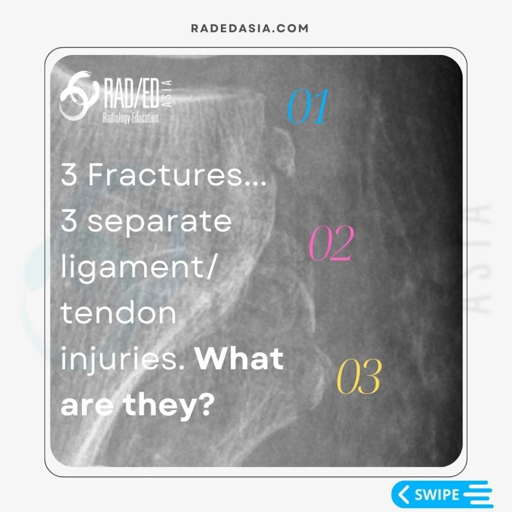
3 LATERAL KNEE FRACTURES SEGOND ARCUATE AND LATERAL FIBULAR FRACTURES XRAY

3 LATERAL KNEE FRACTURE: WHAT THEY ARE AND WHAT THEY MEAN 01: SEGOND FRACTURE • Avulsion Fracture of the lateral tibial plateau.  • Occurs at attachment site of the Antero Lateral Ligament.  • Posterior to IT B insertion site.  • Associated with ACL tears. 02: ARCUATE FRACTURE • Fracture of the Fibula Styloid (tip of …

3 LATERAL KNEE FRACTURES SEGOND ARCUATE AND LATERAL FIBULAR FRACTURES XRAY Leer más »

spine-thoracic-epidural-lipomatosis- radiology-spinal-spine-mri-radedasia

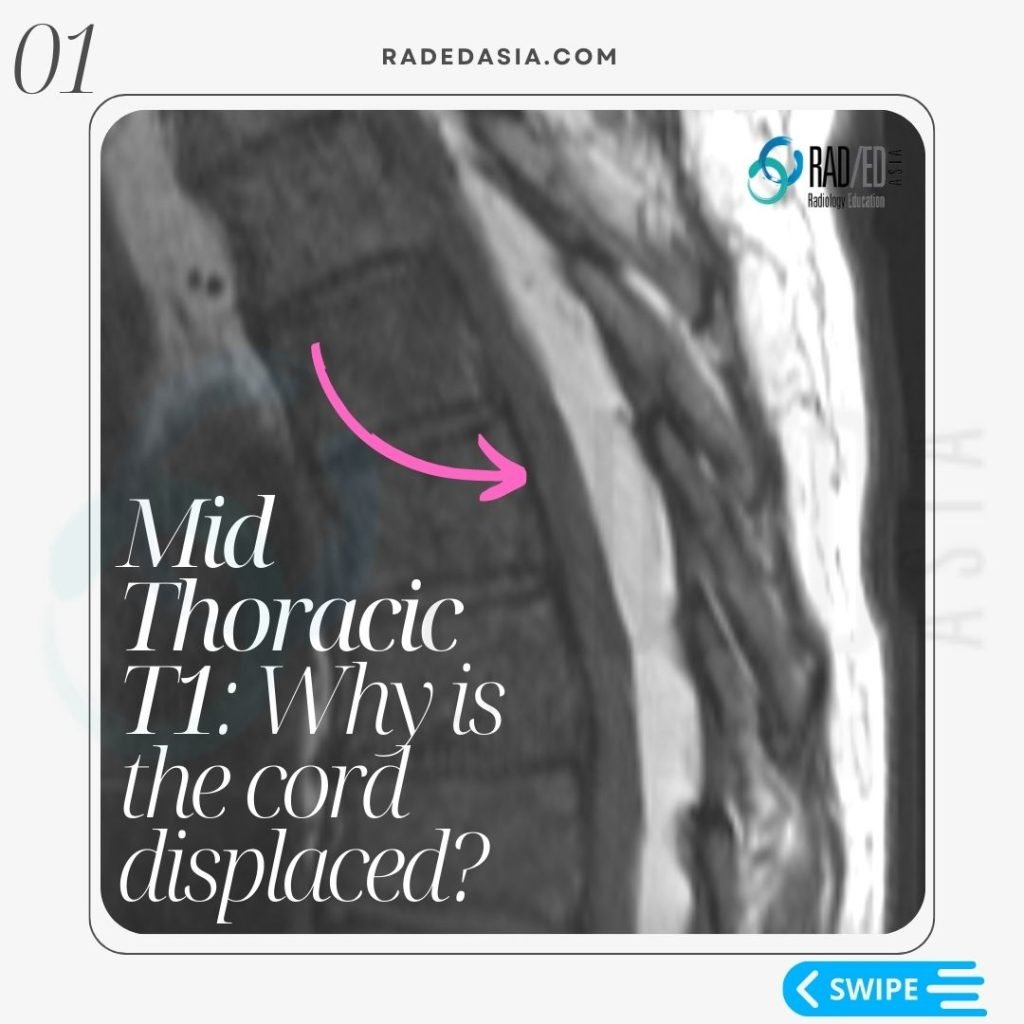
EPIDURAL SPACE INVADERS: SPINE MRI EPIDURAL LIPOMATOSIS PART 2

THORACIC EPIDURAL LIPOMATOSIS RADIOLOGY OVERVIEW Thoracic Epidural lipomatosis is not uncommon to see but has a different appearance to Lumbar Epidural Lipomatosis WHAT IS IT? With thoracic epidural lipomatosis there is excess fat accumulated in the epidural space. WHAT ARE THE CAUSES? There is an increased risk of developing epidural lipomatosis with: Chronic corticosteroid use. …

EPIDURAL SPACE INVADERS: SPINE MRI EPIDURAL LIPOMATOSIS PART 2 Leer más »

meniscal tear radiology mri knee radedasia

MENISCUS TEAR MRI KNEE: MISSING MENISCI WHAT TO LOOK FOR

MENISCUS TEAR MRI KNEE WHAT TO LOOK FOR • In a Normal meniscus the superior and inferior halves should be symmetric. • In all three cases there is localised loss of ” volume” of the meniscus. • This gives the meniscus an asymmetric look. • Asymmetry in a meniscus is always abnormal and we need …

MENISCUS TEAR MRI KNEE: MISSING MENISCI WHAT TO LOOK FOR Leer más »

knee mri bursa suprapatellar plica

KNEE MRI PLICA SUPRAPATELLAR BURSA LIPOHEMARTHROSIS

KNEE MRI PLICA SUPRAPATELLAR BURSA LIPOHEMARTHROSIS NORMAL SUPRA PATELLAR PLICA The normal supra patellar plica is not continuous.  There is a gap in it ( fenestration) which allows fluid to circulate freely in the SP bursa above and below the plica. CONTINUOUS SUPRAPATELLAR PLICA At times the SP Plica is continuous with no gap.  Normally …

KNEE MRI PLICA SUPRAPATELLAR BURSA LIPOHEMARTHROSIS Leer más »

knee infrapatellar plica syndrome radiology mri radedasia

KNEE INFRAPATELLAR PLICA SYNDROME RADIOLOGY MRI (VIDEO)

KNEE INFRAPATELLAR PLICA SYNDROME RADIOLOGY MRI DISCUSSION ON KNEE INFRAPATELLAR PLICA PLICAS IN THE KNEE The infrapatellar plica is one of 4 plicas in the knee: Medial Patella Plica. Suprapatellar Plica. Infrapatellar plica. Lateral Patellar plica. INFRAPATELLAR PLICA ANATOMY Attaches to anterior intercondylar notch and inferior pole patella.   Runs through Hoffa’s Fat Pad.   …

KNEE INFRAPATELLAR PLICA SYNDROME RADIOLOGY MRI (VIDEO) Leer más »

Este sitio está destinado únicamente a profesionales médicos. El uso de este sitio se rige por nuestra Términos de servicio y Declaracion de privacidad que se puede encontrar haciendo clic en los enlaces. Acepte antes de continuar con el sitio web.
error: Clic derecho no disponible.