Ligamento cruzado anterior
This video provides a practical guide to accurately identify small Segond fractures on MRI. We look at 3 findings to look for that will help you make the diagnosis.
![]()
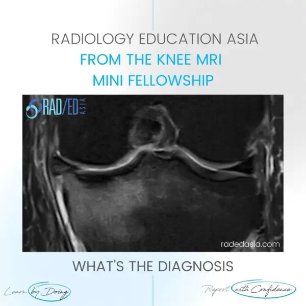
In this video, we’re going to look at How to find small fractures on MRI? Now, it’s difficult on MRI with small fractures, not as easy as CT or even x-ray but there are some clues, we can use to make it a bit easier to make the diagnosis of a small fracture. This video has come from a talk we gave with Hitachi medical systems, Singapore. We need to also look at How do you find a small fracture? because sometimes you may not have the x-ray available to look at or they have, you know usually there isn’t a CT performed. This usually goes from x-ray to MRI so small fractions can be pretty difficult on MRI, and you actually may not see the fragment itself so you have to look for other things that point you towards that being a fracture and a couple of things we can look at. So the first thing you focus on the area of edema, the second bit is to look for localized loss of cortex if we look at this medial tibial plateau first if you follow the cortical line, so cortex is dense bone so it’s black so what you expect to see is a nice black line that is continuous all the way through. In this person who’s got a segond fracture what we see is a nice black line then there’s a bit missing and then here’s the black line again. We can’t see the fracture fragment very well, but the absence of this black line tells you that there is a fracture there so with small fractures where you may not see the fragment this is your clue to there being a fracture. The absence of this black cortical line where in the region where there’s edema okay what about another thing, another thing to look at is to look for a step in the cortex so here is where the fracture fragment has been slightly displaced from the parent bone and the pink arrows pointing to the normal cortex so we have the normal cortical line. Here’s the cortical line above and then you see that they don’t match, there’s a gap there between the two of them. So, the things that we’re really looking for when you’re looking for a small second fracture is to focus on the area of edema, look for localized loss of cortex and then also look for a step for the cortex. You may have one two or three of these, you don’t have to have all of them.
![]()
![]()
A Segond fracture is an avulsion fracture of the lateral tibial plateau at the site of attachment of the Anterior Longitudinal Ligament (ALL). It is strongly associated with an underlying anterior cruciate ligament (ACL) tear.![]()
Key MRI findings include a a step or break in the black cortical line, bony avulsion fragment at the lateral tibial plateau and in the acute stage bone marrow edema.![]()
A Segond fracture typically results from internal rotation and varus stress to the knee. This mechanism puts excessive tension on the lateral capsular ligament, leading to the avulsion at the site of attachment of the Anterior Longitudinal Ligament (ALL).![]()
A Segond fracture involves the lateral aspect of the tibia due to internal rotation and varus stress. A reverse Segond fracture occurs on the medial side of the tibia from external rotation and valgus stress and is associated with posterior cruciate ligament (PCL) and medial meniscus injuries.![]()