
SESAMOID OSTEONECROSIS RADIOLOGY XRAY MRI
OSTEONECROSIS OF THE SESAMOID
- Osteonecrosis of the sesamoids of the 1st metatarsal is not common but important that it’s recognized.
- As it progresses fracture, fragmentation and collapse of the sesamoid can occur.

If we start with Xrays, what do we look for:
- On X-ray look initially for increased bone density/ sclerosis.
- As it progresses fracture, fragmentation and collapse of the sesamoid can occur.

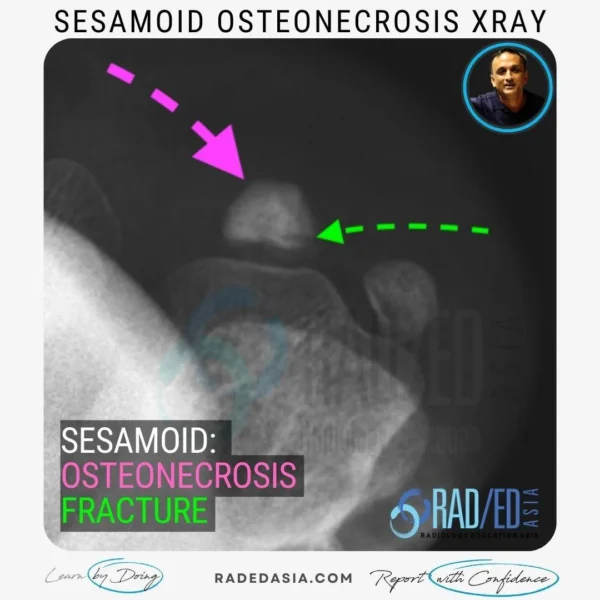
- Sesamoid view demonstrates marked sclerosis of the lateral sesamoid (Pink arrow) with a fracture (Green arrow).
- Compare the degree of sclerosis of the osteonecrotic lateral sesamoid with the normal density of the medial sesamoid.

On CT, what do we look for:
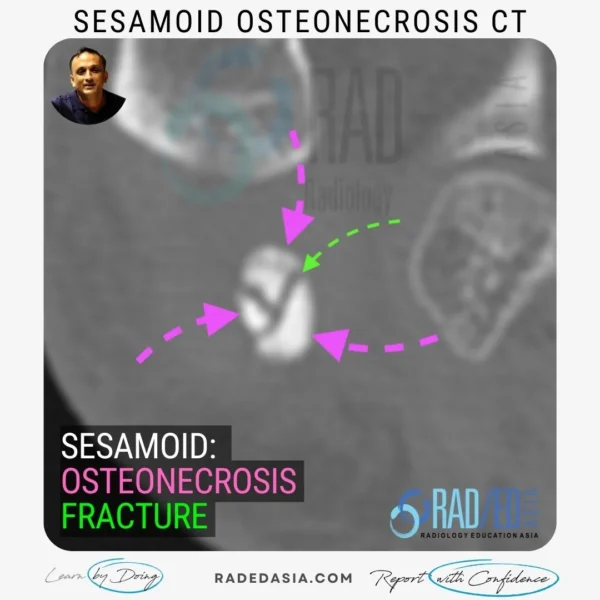
- With osteonecrosis the sesamoid looses its normal lucent central appearance and becomes uniformly hyperdense.
- There may or may not be associated fractures which will be seen as lucent lines.

- Assess for osteonecrosis on the non fat saturated PD or T1 as it’s the fat signal that’s important.
- The normal sesamoid is full of marrow so it will be high signal on PD/ T1.
- On MRI, Osteonecrosis of the sesamoid will demonstrate loss of the normal high marrow signal on PD/ T1.
- It will be low signal on PD/T1.
- PDFS will be low signal with both normal sesamoid and a sesamoid with osteonecrosis.

Osteonecrosis of the sesamoids is important to recognize as it can lead to fracture, fragmentation, and collapse of the sesamoid.

Increased bone density/sclerosis and possible fracture, fragmentation, and collapse of the sesamoid.

Yes, fractures may be seen as lucent lines on X-ray.

The sesamoid will lose its normal lucent central appearance and become uniformly hyperdense. Fractures may or may not be present as lucent lines.

Osteonecrosis of the sesamoid will show loss of the normal high marrow signal on PD/T1 and appear as low signal intensity.

The non-fat saturated PD or T1 sequence is important as the fat signal is crucial.

Learn more about this condition & How best to report it in more detail in our Online Guided ANKLE MRI or FOOT & TOE MRI Mini Fellowship.
More by clicking on the images below.
For all our other current MSK MRI & Spine Imaging
Online Guided Mini Fellowships.
Click on the image below for more information.
- Join our WhatsApp RadEdAsia community for regular educational posts at this link: https://bit.ly/radedasiacommunity
- Get our weekly email with all our educational posts: https://bit.ly/whathappendthisweek

