

OSTEONECROSIS OF THE SESAMOID
- Osteonecrosis of the sesamoids of the 1st metatarsal is not common but important that it’s recognized.
- As it progresses fracture, fragmentation and collapse of the sesamoid can occur.

If we start with Xrays, what do we look for:
- On X-ray look initially for increased bone density/ sclerosis.
- As it progresses fracture, fragmentation and collapse of the sesamoid can occur.
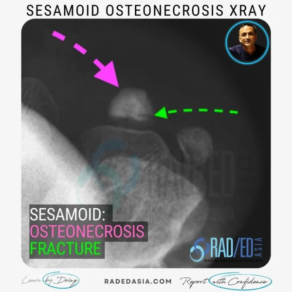
- Sesamoid view demonstrates marked sclerosis of the lateral sesamoid (Pink arrow) with a fracture (Green arrow).
- Compare the degree of sclerosis of the osteonecrotic lateral sesamoid with the normal density of the medial sesamoid.
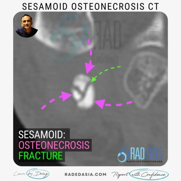
On CT, what do we look for:
- With osteonecrosis the sesamoid looses its normal lucent central appearance and becomes uniformly hyperdense.
- There may or may not be associated fractures which will be seen as lucent lines.
- Assess for osteonecrosis on the non fat saturated PD or T1 as it’s the fat signal that’s important.
- The normal sesamoid is full of marrow so it will be high signal on PD/ T1.
- On MRI, Osteonecrosis of the sesamoid will demonstrate loss of the normal high marrow signal on PD/ T1.
- It will be low signal on PD/T1.
- PDFS will be low signal with both normal sesamoid and a sesamoid with osteonecrosis.
![]()
Osteonecrosis of the sesamoids is important to recognize as it can lead to fracture, fragmentation, and collapse of the sesamoid.
![]()
Increased bone density/sclerosis and possible fracture, fragmentation, and collapse of the sesamoid.
![]()
Yes, fractures may be seen as lucent lines on X-ray.
![]()
The sesamoid will lose its normal lucent central appearance and become uniformly hyperdense. Fractures may or may not be present as lucent lines.
![]()
Osteonecrosis of the sesamoid will show loss of the normal high marrow signal on PD/T1 and appear as low signal intensity.
![]()
The non-fat saturated PD or T1 sequence is important as the fat signal is crucial.
![]()