There are a number of appearances of periosteal reaction on MRI secondary to trauma that we can see and report.
The Histology...Yes Histology!: It helps to have a simple understanding of the histology of the periosteum and the following is from a nice review article in Skeletal Radiology by Jerry Dwek: The periosteum: what is it, where is it, and what mimics it in its absence? Skeletal Radiol (2010) 39:319–323
"Periosteum can be thought of as consisting of two distinct layers, an outer fibrous layer and an inner layer ( cambium) that has significant osteoblastic potential. "
"After a fracture, a hematoma forms, which is stabilized by the surrounding soft tissues and a short time later by a reconstituted (if it is indeed torn) fibrous layer of the periosteum. Cells from the inner cambium layer proliferate and differentiate. At the periphery of the fracture, where good vascularity is present, the inner periosteal layer lays down a collar of bone by the process of membranous ossification. Nearer to the fracture site, the cambium produces a mass of cartilage about the fracture site. Then, by a process of endochondral ossification ... the cartilage mass is ossified. "
Periosteum is not seen separate to underlying cortex as both are black on MRI. What you see is a thick rim of black cortex which includes the periosteum.

Image Above PD and PDFS: Normal Cortex and periosteum are indistinguishable (Blue arrows) as both are black in signal. No fluid or oedema at superficial margin of cortex.
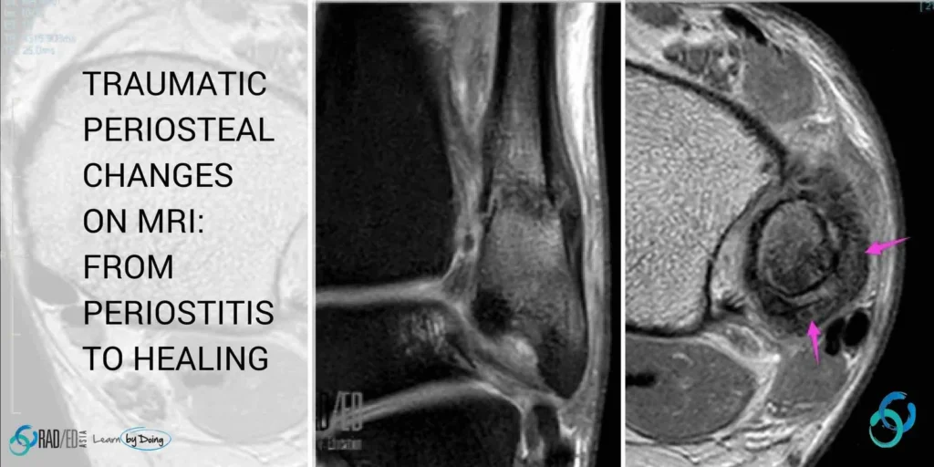
Periostitis is inflammatory change of the periosteum and in the context of trauma, is an early change of stress response. There is no periosteal new bone formation as yet, but cortical margin becomes ill defined, and oedema is present in the soft tissues adjacent to the periosteum.
 Image above: Contrast the normal sharp black line of cortex and periosteum (Pink arrows) with the localized area of periostitis with ill definition of the cortical margin (blue arrows) and overlying soft tissue oedema (Yellow arrows).
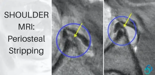
Normally, where ligaments attach to bone they are incorporated into and covered by periosteum. So in an acute injury the ligament attachment site can tear by stripping the periosteum with it. Look for a thin extra black line overlying cortex (periosteum) and high PD signal tracking along the cortex which is oedema and hemorrhage.
 Image Above: Distal superficial MCL tear and avulsion has resulted in stripping of the periosteum ( thin black line blue arrows) at its attachment. High PD signal is oedema and hemorrhage.
Similar to above. Look for a thin extra black line overlying cortex (periosteum) and high PD signal tracking along the cortex which is oedema and hemorrhage.

 Image Above: Posterior malleolar fracture (not shown) with black line of secondary periosteal new bone formation (yellow arrows) and oedema (blue arrows). Purple arrows underlying cortex.
Callus formation is at the level of the fracture and is much thicker and of an intermediate to higher signal than periosteum.
 Image Above: Distal fibular fracture (blue arrow) with thick intermediate signal callus formation (pink arrows) at level of fracture and thin periosteal new bone formation above the fracture site (yellow arrows). Axial image through level of callus.
Ossification of callus and the periosteal new bone results in a thick cortical margin which is of low signal.

Image above: First image acute metatarsal stress fracture with periosteal new bone formation (Yellow arrows) and callus (pink arrow). Second image fracture healing with ossification of callus and periosteal new bone resulting in a thick medial cortex (Blue arrows).
Link to article:


ANKLE
ANKLE