brain

CEREBELLAR INFARCTS: HOW NOT TO MISS THEM

The Problem: Acute cerebellar infarcts are easy to see on diffusion imaging, but small chronic infarcts in the cerebellum are often missed because they are confused with normal cerebellar sulci. How do you differentiate small chronic cerebellar infracts from normal sulci? The Answer: In the Cerebellar Hemispheres: Normal: Chronic Infarct: Infarcts usually run perpendicular to the …

CEREBELLAR INFARCTS: HOW NOT TO MISS THEM Read More »

tumefactive-cerebral-amyloid-mri

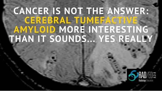
CEREBRAL TUMEFACTIVE AMYLOID: CANCER IS NOT THE ANSWER

INTRO: Cerebral tumefactive amyloid is a rare mass like form of cerebral amyloid. PATHOLOGY: Caused by vasculitis +/- perivasculitis which is an autoimmune response to the amyloid deposition. KEY IMAGING POINTS: MRI findings similar to a glioma/ treated lymphoma with T2 hyper intensity and mass effect. Can demonstrate leptomeningeal enhancement over the region of abnormality, however …

CEREBRAL TUMEFACTIVE AMYLOID: CANCER IS NOT THE ANSWER Read More »

SWI OR T2*: WHICH MRI SEQUENCE TO USE IN THE DETECTION OF CEREBRAL MICROBLEEDS?

Quick Journal Review: Should you use T2* or SWI sequences to detect cerebral microbleeds.Which sequence is more sensitive? Journal Article: SWI or T2*: Which MRI Sequence to Use in the Detection of Cerebral Microbleeds? The Karolinska Imaging Dementia Study S. Shams et al AJNR 36:1089–95 Jun 2015 Intro: Different sequences can be used to detect …

SWI OR T2*: WHICH MRI SEQUENCE TO USE IN THE DETECTION OF CEREBRAL MICROBLEEDS? Read More »

This site is intended for Medical Professionals only. Use of this site is governed by our Terms of Service and Privacy Statement which can be found by clicking on the links. Please accept before proceeding to the website.
error: Right Click not available.