This site is intended for Medical Professions only. Use of this site is governed by our Terms of Service and Privacy Statement which can be found by clicking on the links. Please accept before proceeding to the website.

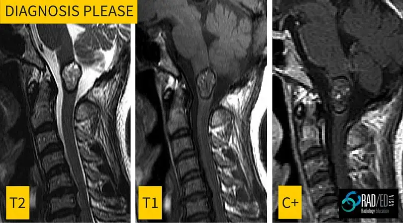
Radiology Education Diagnosis Please: Spinal cord lesion MRI
This is the first in a regular series of posts on cases of interest.
Incidental finding in a 40yo male. Image above.
ANSWER
Cavernomas of the spinal cord have a very specific appearance on MRI with the following features
- Lobulated well defined lesion mixed signal T1 and T2
- No significant enhancement
- Low signal rim of hemosiderin on all sequences represents slow leak of blood products
- Mass effect but no oedema unless there has been acute bleeding
- Blooming on Gradient Echo sequence
Upcoming MRI Spine Workstation Workshop in Singapore 1st October 2016. More information at this link mrispinesg


