scapula normal variant keats shoulder mri radiology radedasia

NORMAL SCAPULA VARIANT SHOULDER MRI RADIOLOGY (VIDEO)

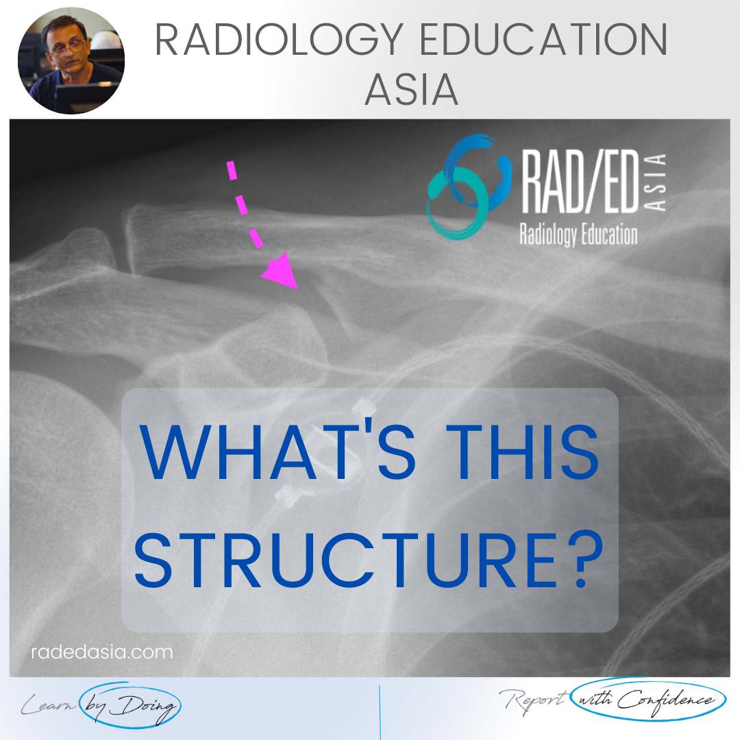
Well corticated bridge of bone (Pink arrows) that lies on the superior margin of the scapula with a bone defect inferior to it.

  • This is a normal variant. Its described in Keats as a Clasp Like scapular margin that’s a result of bone being developmentally absent below it.


  • This results in a pseudoforamen at the superior scapular margin.

If your Browser is blocking the video, Please view it on our YouTube Channel HERE.

Learn more about SHOULDER Imaging in our ONLINE Guided MRI SHOULDER Mini-Fellowship.


More by clicking on the images below.

coracoclavicular joint shoulder normal variant xray radedasia
SHOULDER
error: Content is protected !!

Join our Newsletter

Stay tuned on new
Mini-Fellowships launches and learnings

Mini-Fellowships

Better Than Conferences

Join our WhatsApp Community

We help radiologists learn
by doing and report with confidence

This site is intended for Medical Professions only. Use of this site is governed by our Terms of Service and Privacy Statement which can be found by clicking on the links. Please accept before proceeding to the website.