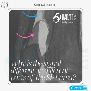
patella plica mri radiology syndrome knee medial radedasia

KNEE PLICA MRI MEDIAL PATELLAR PLICA RADIOLOGY

  • The medial patella plica is a developmental synovial membrane remnant.
 
  • Can be seen in up to 30% of knees.
  • It can be completely asymptomatic but can extend between the patella and trochlea and be compressed and cause pain. This is seen in so called Patella Plica Syndrome.


  • May also have chondromalacia in the adjacent patella and trochlea facet.

  • A normal plica as shown is well defined and low signal.


  • It is found in the superomedial aspect of the joint and is best seen on axial scans.


  • With compression it will be thickened and ill defined and increase in signal.

patella-plica-mri-radiology-syndrome-knee-medial-radedasia.jpgmri-patella-plica-radiology-syndrome-knee-medial-radedasia.

Learn more about KNEE Imaging in our ONLINE or ONSITE
Guided MRI KNEE Mini-Fellowship.


More by clicking on the images below.

knee mri bursa suprapatellar plica
KNEE
knee-infrapatellar-plica-syndrome-radiology-mri-radedasia-1
KNEE

#radiology #radedasia #mri #kneemri #msk #mriknee #mskmri #radiologyeducation #radiologycases #radiologist #rads #radiologystudent #radiologycme #radiologycpd #medicalimaging #imaging #radcme #rheumatology #arthritis #rheumatologist #sportsmed #orthopaedic #physio #physiotherapist #kneeplica #plica #medialpatellaplica

error: Content is protected !!

Join our Newsletter

Stay tuned on new
Mini-Fellowships launches and learnings

Mini-Fellowships

Better Than Conferences

Join our WhatsApp Community

We help radiologists learn
by doing and report with confidence

This site is intended for Medical Professions only. Use of this site is governed by our Terms of Service and Privacy Statement which can be found by clicking on the links. Please accept before proceeding to the website.