acl-pcl-mucoid-degeneration-mri-findings-radedasia

Stay tuned on new Fellowships and learnings

ACL & PCL MUCOID DEGENERATION MRI FINDINGS

MRI FINDINGS IN ACL AND PCL MUCOID DEGENERATION

ACL AND PCL MUCOID DEGENERATION ON MRI


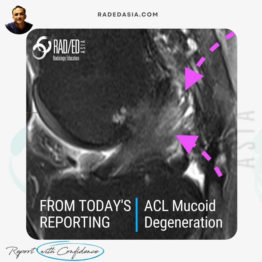
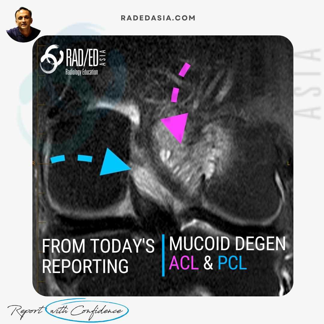
Mucoid degeneration can affect both the anterior cruciate ligament (ACL) and posterior cruciate ligament (PCL). Mucoid degeneration is often confused with ACL tears, and this post looks at the main MRI features of ACL and PCL mucoid degeneration that will help to differentiate from tears.

radiology-education-asia-radedasia
 

MRI FEATURES OF ACL & PCL MUCOID DEGENERATION

acl-mucoid-degeneration-celery-stalk-sign-mri-radedasia

WHAT ARE THE FINDINGS?

  • The ACL is enlarged and hyperintense but there is continuity of fibres and normal alignment.

  • The enlargement is more prominent proximally giving it the appearance of a celery stalk (celery stalk sign of ACL mucoid degeneration).


SUMMARY:

This is a characteristic appearance of ACL mucoid degeneration. Look for, 

  • Expanded hyperintense ACL.
     
  • Fibres are continuous.
     
  • Alignment of fibres is normal.

radiology-education-asia-radedasia

acl-mucoid-degeneration-cysts-mri-radedasia

  • Intra ligamentous cysts are commonly found in mucoid degeneration of the ACL.

  • Size can be variable from small cysts like in this image to larger ones we will see next.
  • Look for T2 hyperintense cystic changes in the ACL.


radiology-education-asia-radedasia

acl-mucoid-degeneration-intraosseous-cysts-mri-radedasia

  • Cysts can extend from the ACL into the bony attachments at the tibia or femur.
  • Look for cystic change of variable size at the femur and tibia at the attachment site.
  • Intraligamentous cysts can also extend into the adjacent soft tissue.

radiology-education-asia-radedasia

knee ligament mucoid degen acl pcl mcl radiology radedasia

  • It’s not just the ACL that undergoes mucoid degeneration.
  • The PCL can also be involved but it’s less common.
  • Mucoid degeneration of the PCL can be in conjunction with the ACL or on its own.
  • Look for the same findings discussed for the ACL to make the diagnosis.

radiology-education-asia-radedasia

  • Ligament thickening.
  • “Celery stalk” sign.
  • Increased T2 Signal intensity.
  • Preserved fibers which are in continuity.
  • Normal alignment of the ACL or PCL.
  • Intra ligamentous cysts of variable size.
  • Intra osseous extension of the cysts.

radiology-education-asia-radedasia

Our CPD & Learning Partners

OUR POPULAR POSTS ON KNEE MENISCUS: CLICK ON THE IMAGES BELOW

Stay tuned on new Fellowships and learnings

error: Content is protected !!

Join our Newsletter

Stay tuned on new
Mini-Fellowships launches and learnings

Mini-Fellowships

Better Than Conferences

Join our WhatsApp Community

We help radiologists learn
by doing and report with confidence

This site is intended for Medical Professions only. Use of this site is governed by our Terms of Service and Privacy Statement which can be found by clicking on the links. Please accept before proceeding to the website.