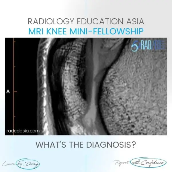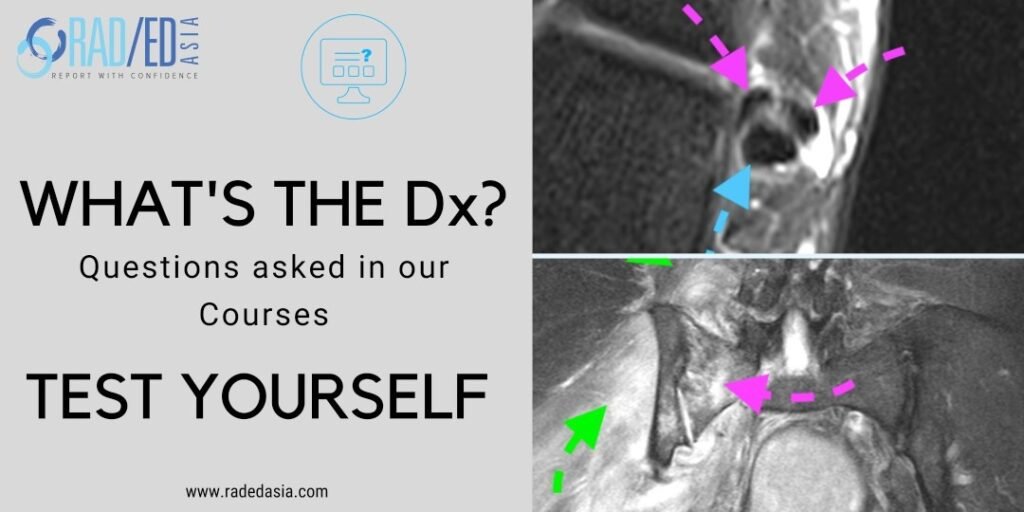
ACL TEAR MRI KNEE FINDINGS CORONAL IMAGES
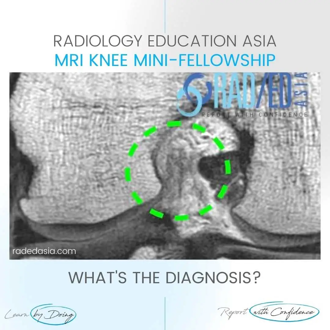
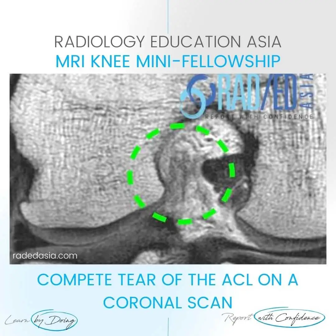
- This is a compete tear of the ACL on a coronal scan.
- The ACL (Green circle) is markedly thinned, ill defined, hyperintense and irregular.
- Compare with the normal low signal and good definition of the adjacent PCL.
- The ACL should have a similar appearance to the PCL.

ACL (Green circle) is markedly thinned, ill defined, hyperintense and irregular.

