mri acl tear pivot shift injury bone contusion pattern osteochondral fracture knee

ACL TEAR PIVOT SHIFT INJURY BONE CONTUSION PATTERN OSTEOCHONDRAL FRACTURE MRI KNEE

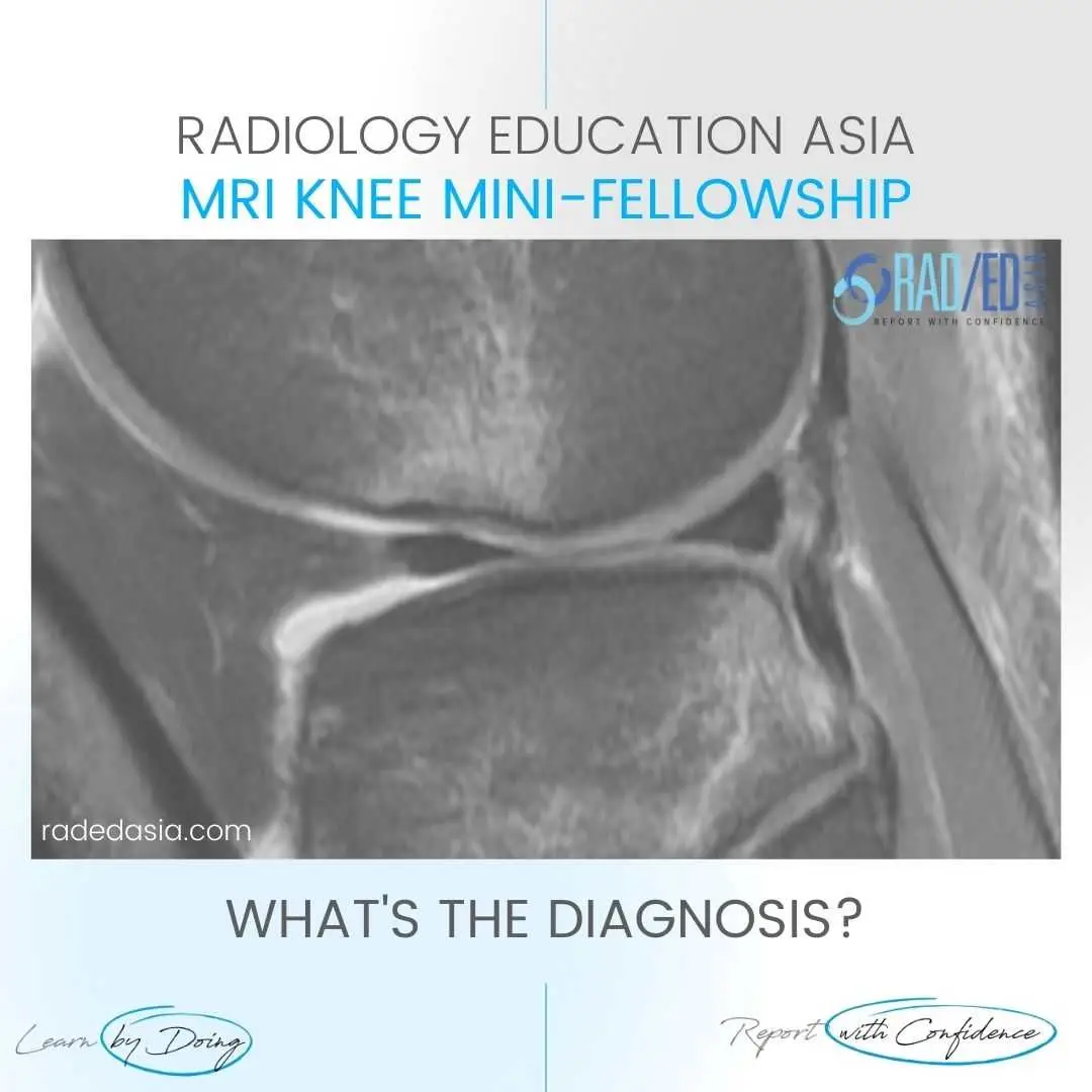
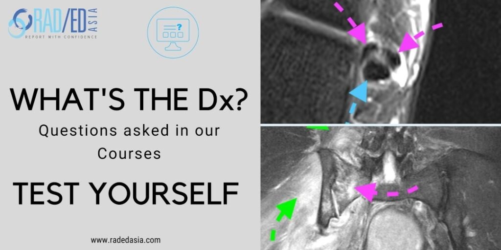
  • Pivot shift bone bruising of the lateral femoral condyle and an osteochondral depression fracture secondary to an ACL tear.


  • Characteristic bone contusion pattern seen with complete ACL tear.


  • The osteochondral fracture is seen with the flattening of the cortical margin with underlying bone marrow oedema.

  • Bone bruising (Pink arrows) Lateral Femoral Condyle and posterior aspect Lateral tibial plateau.


  • Osteochondral depression fracture lateral femoral condyle (Green arrow).

mri-acl-tear-pivot-shift-injury-bone-contusion-pattern-osteochondral-fracture-knee.jpg

#radiology #radiologist #radiologia #mri #kneemri #mskmri #medicalimaging #imaging #futureradiologist #radiologyresident #frcr #foamrad #ortho #ct #emergencydepartment #orthopaedics #radiologylife #trauma #radiologystudent #mskrad #diagnosticimaging #mskradiology #radedasia #radiologyeducation #radiologycases #radiologycme #rheumatology #arthritis #rheumatologist #degenerativedisease #painphysician #chiropractic #physiotherapy #sportsmed

error: Content is protected !!

Join our Newsletter

Stay tuned on new
Mini-Fellowships launches and learnings

Mini-Fellowships

Better Than Conferences

Join our WhatsApp Community

We help radiologists learn
by doing and report with confidence

This site is intended for Medical Professions only. Use of this site is governed by our Terms of Service and Privacy Statement which can be found by clicking on the links. Please accept before proceeding to the website.