- 7th March 2026
SGD$695.00
ANKYLOSING SPONDYLITIS DAGGER SIGN SPINE IMAGING: WHAT DOES IT MEAN

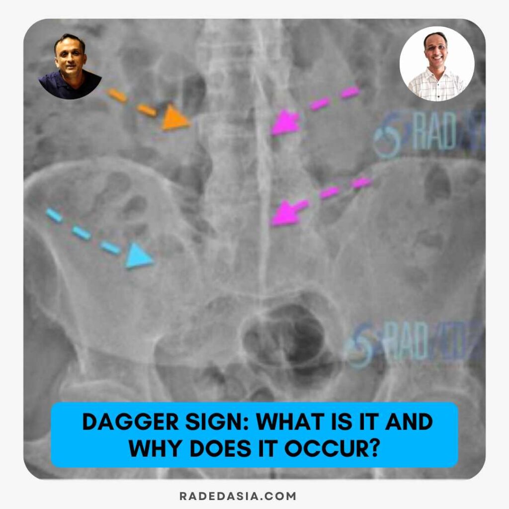
ANKYLOSING SPONDYLITIS DAGGER SIGN SPINE IMAGING
WHAT IS THE DAGGER SIGN
WHAT DO I LOOK FOR
WHY DOES IT OCCUR
WHAT'S THE LIGAMENT ANATOMY
- INTERSPINOUS LIGAMENT (Pink shading)
- Attaches to the cranial and caudal spinous process at each level.
- But also has attachment to the lamina and ligamentum flavum.
- SUPRASPINOUS LIGAMENT (Blue shading).
- Attaches between the tips of the spinous processes (Thoracic and lumbar).
- Above C7 it is free of the spinous processes and attaches to the occiput as the ligamentum nuchae.

WHAT DO THE LIGAMENTS LOOK LIKE ON DISSECTION
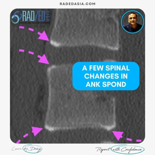
WHAT'S THE PATHOLOGY
HOW TO REPORT

READ MORE
Read article: “A Comprehensive Assessment of Hip Damage in Ankylosing Spondylitis, Especially Early Features”, Read HERE
 This post has been made in conjunction with Dr Joe Thomas, a Senior Consultant Rheumatologist at Aster Hospital in Kochi India, who has a vast amount of clinical experience. He also has a very strong interest in Imaging of Arthropathies and has joined us to bring a clinical perspective to the imaging and to advise on what rheumatologists want when we report their referrals.
 - Join our WhatsApp RadEdAsia community for regular educational posts at this link: https://bit.ly/radedasiacommunity
- Get our weekly email with all our educational posts: https://bit.ly/whathappendthisweek
- 7th March 2026
SGD$695.00


 SP Spinous Process, SSL Supraspinous Ligament FJC Facet Joint Capsule.


