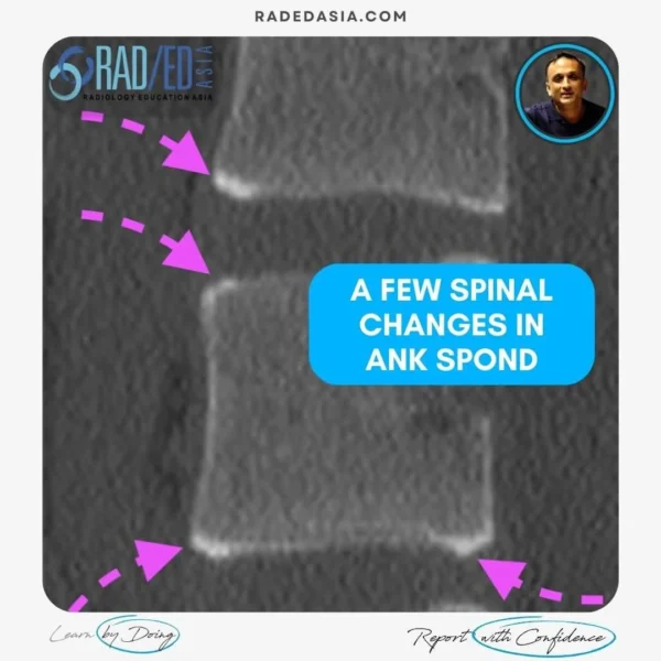
Ankylosing Spondylitis
In ankylosing spondylitis, what is the dagger sign? In this post we look at how to recognize it and he Anatomy and Pathology that helps explain what the Dagger sign is.
![]()
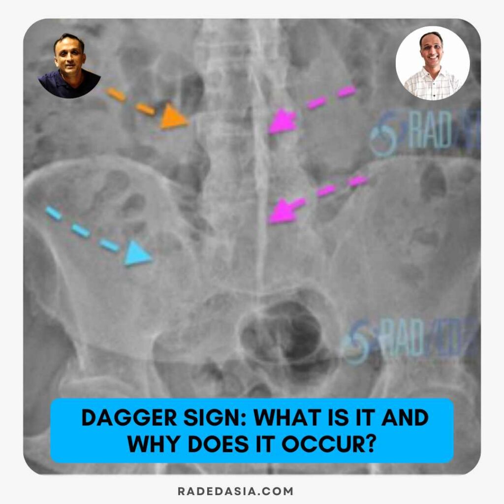
In Ankylosing spondylitis, the dagger sign refers to a solid central line of bone along in AP spine view.![]()
Linear density in the midline connecting the spinous processes (Pink arrows).

Image Above: Dagger sign (Pink arrows). Also, SIJ fusion (Blue arrow) and Bamboo spine (Orange arrow).
It’s due to ossification of the interspinous and supraspinous ligaments that extend between the spinous processes.![]()

 SP Spinous Process, SSL Supraspinous Ligament FJC Facet Joint Capsule.
Image from 2019 Asian Journal of Neurosurgery Anatomical and Biomechanical Study of the Lumbar Interspinous Ligament.![]()
Caused by enthesitis of the ligamentous attachments to the spinous process followed eventually by ossification.![]()
The dagger sign is visualized on frontal x-ray and coronal CT.![]()

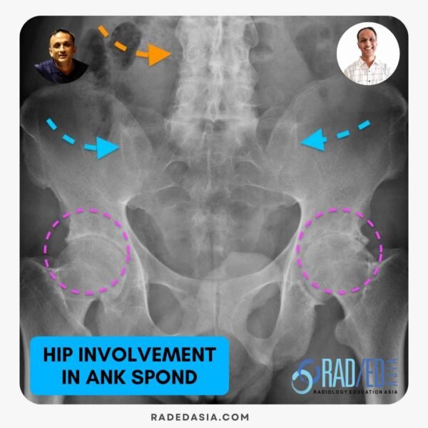
Read article: “A Comprehensive Assessment of Hip Damage in Ankylosing Spondylitis, Especially Early Features”, Read HERE
![]()
 This post has been made in conjunction with Dr Joe Thomas, a Senior Consultant Rheumatologist at Aster Hospital in Kochi India, who has a vast amount of clinical experience. He also has a very strong interest in Imaging of Arthropathies and has joined us to bring a clinical perspective to the imaging and to advise on what rheumatologists want when we report their referrals.