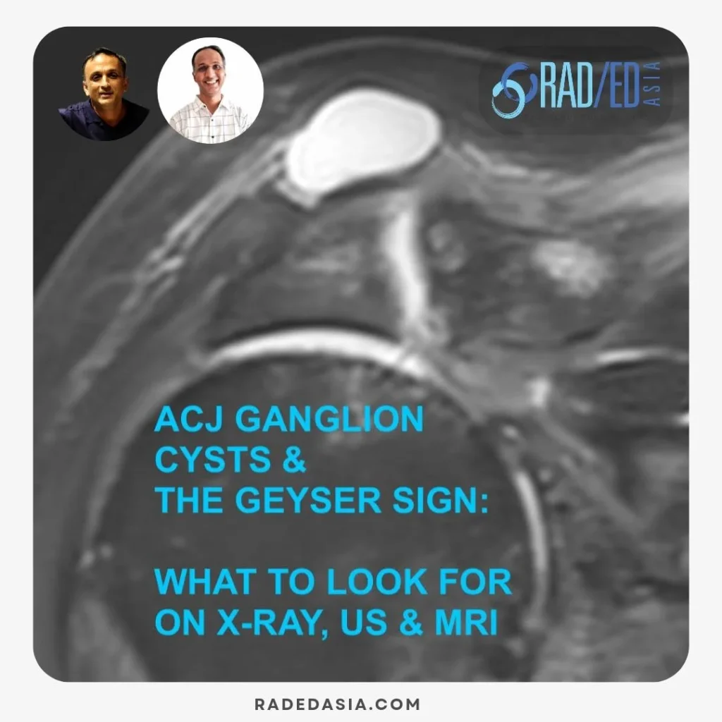
Ravi

acromial spur mri shoulder radiology education asia

ACROMION / ACROMIAL SPURS ON MRI: WHERE TO LOOK AND WHAT TO LOOK FOR

ACROMION SPURS ON MRI: 3 APPEARANCES ON MRI   Acromial / Acromion spurs can be quite small and difficult to visualize on MRI.   They are enthesophytes that arise from traction at the attachment sites of the Coraco Acromial Ligament and Deltoid.   This post looks at the three locations and most common appearances of

ACROMION / ACROMIAL SPURS ON MRI: WHERE TO LOOK AND WHAT TO LOOK FOR Read More »

mri knee fat pad impingement radiology education asia

KNEE MRI FAT PAD IMPINGEMENT: WHERE TO LOOK & WHAT TO LOOK FOR

Stay tuned on new Fellowships and learnings Subscribe The Dx / Knee KNEE FAT PAD IMPINGEMENT MRI APPEARANCE MRI OF FAT PAD IMPINGEMENT AROUND THE KNEE Knee fat pad impingement syndromes are characterized by pain and inflammation of the fat pads surrounding the knee joint and can significantly impact knee function.   This blog post

KNEE MRI FAT PAD IMPINGEMENT: WHERE TO LOOK & WHAT TO LOOK FOR Read More »

de quervain's tenosynovitis wrist ultrasound how to scan radiology radedasia

DE QUERVAIN’S TENOSYNOVITIS: HOW TO SCAN ON ULTRASOUND (VIDEO)

DE QUERVAIN'S TENOSYNOVITIS: HOW TO SCAN AND INJECT WRIST COMPARTMENT ONE ON ULTRASOUND DE QUERVAIN’S TENOSYNOVITIS: HOW TO SCAN AND INJECT ON ULTRASOUND De Quervain’s tenosynovitis is a stenosing tenosynovitis involving the  retinaculum of the first dorsal compartment of the wrist, with involvement of abductor pollicis longus (APL) and extensor pollicis brevis (EPB) tendons and

DE QUERVAIN’S TENOSYNOVITIS: HOW TO SCAN ON ULTRASOUND (VIDEO) Read More »

cppd pseudogout chondrocalcinosis knee gout radiology xray

CPPD CHONDROCALCINOSIS PSEUDOGOUT KNEE RADIOLOGY (VIDEO)

CPPD PSEUDOGOUT KNEE XRAY: WHICH STRUCTURES ARE INVOLVED CPPD PSEUDOGOUT KNEE XRAY   Calcium pyrophosphate deposition (CPPD) is a common chronic arthropathy caused by the presence of calcium pyrophosphate (CPP) deposits in joint and surrounding tissues. Common arthropathy which increases with age. It can occur in any joint but the knee is a very common

CPPD CHONDROCALCINOSIS PSEUDOGOUT KNEE RADIOLOGY (VIDEO) Read More »

weight bearing x-rays for lisfranc joint injury instability fracture radedasia

WEIGHT BEARING X-RAYS FOR LISFRANC JOINT INJURY INSTABILITY & FRACTURE (VIDEO)

THE MAIN POINTS TO NOTE OVERVIEW WEIGHT BEARING X-RAYS FOR LISFRANC JOINT INJURY INSTABILITY & FRACTURE Early diagnosis of Lisfranc instability is crucial to prevent premature OA in the mid foot.   Subtle instability patterns can be very difficult to identify on xrays.   Plain radiographs may not effectively identify instability at the LisFranc Joint,

WEIGHT BEARING X-RAYS FOR LISFRANC JOINT INJURY INSTABILITY & FRACTURE (VIDEO) Read More »

intersection syndrome wrist ultrasound radiology how to scan radedasia

ULTRASOUND WRIST INTERSECTION SYNDROME HOW TO SCAN & WHAT TO LOOK FOR (VIDEO)

ULTRASOUND WRIST INTERSECTION SYNDROME ULTRASOUND WRIST INTERSECTION SYNDROME HOW TO SCAN & WHAT TO LOOK FOR How and where do you assess on wrist ultrasound for intersection syndrome. In this video by Dr Rajendra Sahoo, he first looks at how and where to scan for De Quervain’s and then how and where to scan for

ULTRASOUND WRIST INTERSECTION SYNDROME HOW TO SCAN & WHAT TO LOOK FOR (VIDEO) Read More »

ct mri facet cysts mri spine how to mri facet cysts spine learn spine mri online radiology education asia

LUMBAR SPINE FACET JOINT SYNOVIAL CYST RADIOLOGY: MRI AND CT APPEARANCE (VIDEO)

LUMBAR SPINE FACET JOINT SYNOVIAL CYSTS MRI AND CT Today’s post is a video on MRI of the Spine and Facet Joints and in particular the MRI and CT appearance of Lumbar Spine Facet Joint Synovial Cysts. This approx 5 minute video below looks at What do spine facet joint cysts look like on MRI

LUMBAR SPINE FACET JOINT SYNOVIAL CYST RADIOLOGY: MRI AND CT APPEARANCE (VIDEO) Read More »

freiberg disease mri radiology toe radedasia

FREIBERG DISEASE SUBCHONDRAL FRACTURE INFRACTION OSTEOCHONDROSIS (VIDEO)

FREIBERG DISEASE / SUBCHONDRAL FRACTURE INFRACTION / OSTEOCHONDROSIS DISCUSSION FREIBERG DISEASE / SUBCHONDRAL FRACTURE INFRACTION / OSTEOCHONDROSIS Extensive oedema in the head of the 2nd metatarsal with slight flattening of the cortical margin is characteristic of what’s called Freiberg’s Disease in adolescents and Subchondral Fracture in adults. Freiberg disease occurs in adolescents with an unfused

FREIBERG DISEASE SUBCHONDRAL FRACTURE INFRACTION OSTEOCHONDROSIS (VIDEO) Read More »

ultrasound-ankle-foot-baxter's-nerve-inferior-calcaneal-nerve-entrapment-radiology-radedasia

BAXTER’S NERVE ULTRASOUND HOW TO FIND IT (VIDEO)

BAXTER'S NERVE ULTRASOUND Dr RAJENDRA SAHOO Dr Rajendra Sahoo is a senior consultant Pain Physician and Anesthetist who is Fellowship trained in Ultrasound guided Pain Management. We will be collaborating with Dr Sahoo to bring more MSK and Spine Ultrasound learning. BAXTER’S NERVE ULTRASOUND: HOW TO FIND IT How do you find the Baxter’s nerve

BAXTER’S NERVE ULTRASOUND HOW TO FIND IT (VIDEO) Read More »

mri plantar fascia tear fasciitis plantar fascia anatomy heel pain mri mri online course radedasia

MRI PLANTAR FASCIA ANATOMY: WHAT ARE THE 3 BANDS OF THE PLANTAR FASCIA (VIDEO)

MRI PLANTAR FASCIA ANATOMY MRI PLANTAR FASCIA ANATOMY: NORMAL BANDS Today’s post is a video on MRI of the Plantar Fascia Anatomy. We are looking at how to identify the 3 bands of the Plantar Fascia and which band is most important in Plantar Fasciitis. If you find the video helpful, please subscribe to the

MRI PLANTAR FASCIA ANATOMY: WHAT ARE THE 3 BANDS OF THE PLANTAR FASCIA (VIDEO) Read More »

error: Content is protected !!

Join our Newsletter

Stay tuned on new
Mini-Fellowships launches and learnings

Mini-Fellowships

Better Than Conferences

Join our WhatsApp Community

We help radiologists learn
by doing and report with confidence

This site is intended for Medical Professions only. Use of this site is governed by our Terms of Service and Privacy Statement which can be found by clicking on the links. Please accept before proceeding to the website.