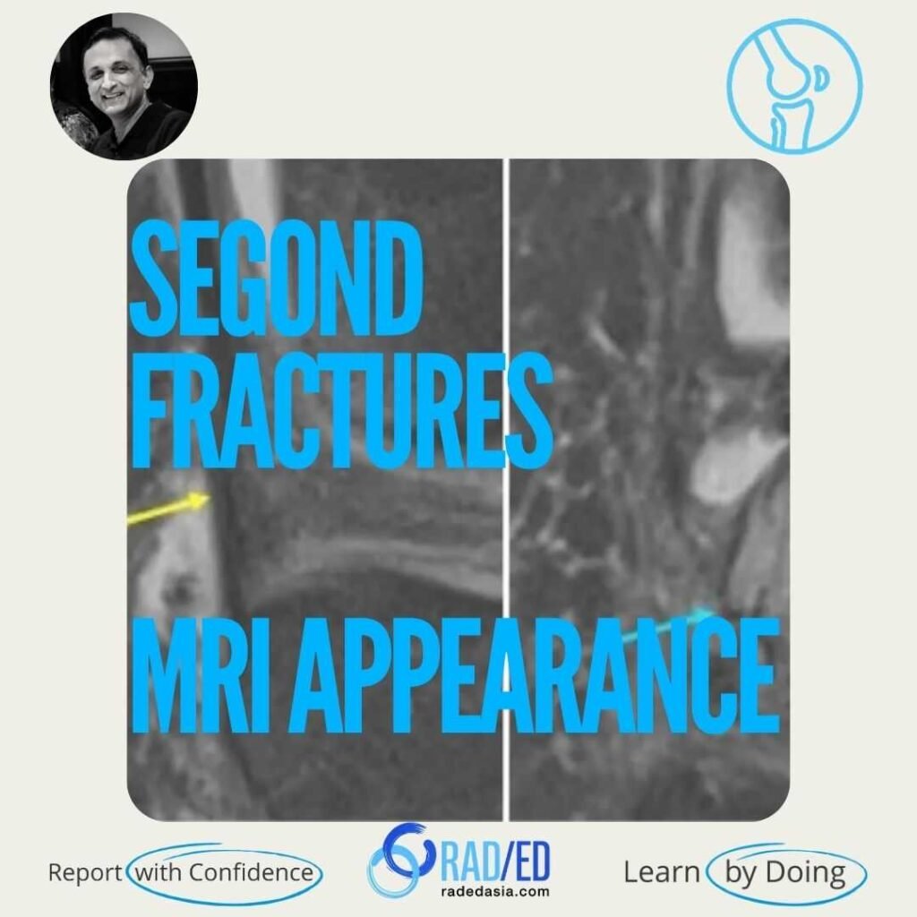
Knee MRI demonstrating the MRI appearance of a Segond fracture, a classic avulsion injury associated with anterior cruciate ligament tears.

Stay tuned on new Fellowships and learnings

MRI SEGOND FRACTURE: WHAT TO LOOK FOR

SEGOND FRACTURE: WHAT TO LOOK FOR ON MRI & X-RAY

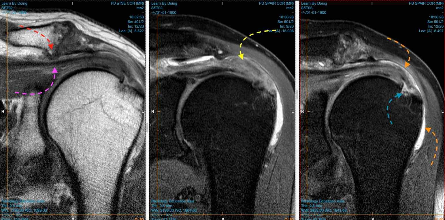
Segond Fractures are fractures of the lateral tibial plateau often associated with ACL tears. It’s thought to be related to the tibial insertion of the anterolateral ligament. However, this ligament is not distinctly identified on MRI, and the best landmark is to look immediately posterior to the ITB insertion.

radiology-education-asia-radedasia
 

Lateral tibial plateau, just posterior to the attachment of the ilio-tibial band.
radiology-education-asia-radedasia

It’s thought to be related to the tibial insertion of the anterolateral ligament. However, this ligament is not distinctly identified on MRI, and the best landmark is to look immediately posterior to the ITB insertion.
radiology-education-asia-radedasia

Small avulsion/ flake type fracture of the lateral tibial plateau.

mri-knee-segond-fracture-radiology-education-asia
radiology-education-asia-radedasia

  • Bone marrow oedema immediately posterior to the ITB insertion. May only be seen as an area of bone marrow oedema rather than the classic fracture fragment seen on x-ray.

  • Bone fragment or loss of cortical margin immediately posterior to the ITB.

mri-knee-segond-fracture-xray-radiology-education-asia

Image Above: X-ray shows fracture fragment (blue arrow)) adjacent to lateral tibial plateau. ITB (yellow arrow) is intact and normal. MRI third image Bone marrow oedema (blue arrow) low signal fracture line seen immediately posterior to ITB insertion.


radiology-education-asia-radedasia

mri-knee-segond-fracture-fragment-radiology-education-asia

Image Above: Segond Fracture normal ITB insertion (yellow arrow). Fracture of the lateral cortex (blue arrows) posterior to ITB insertion.


radiology-education-asia-radedasia

TEST YOURSELF ON SOME COMMON & FREQUENTLY ASKED QUESTIONS

 A Segond fracture is an avulsion fracture of the lateral tibial plateau at the site of attachment of the Anterior Longitudinal Ligament (ALL). It is strongly associated with an underlying anterior cruciate ligament (ACL) tear.
radiology-education-asia-radedasia

Key MRI findings include a a step or break in the black cortical line, bony avulsion fragment at the lateral tibial plateau and in the acute stage bone marrow edema.
radiology-education-asia-radedasia

A Segond fracture typically results from internal rotation and varus stress to the knee. This mechanism puts excessive tension on the lateral capsular ligament, leading to the avulsion at the site of attachment of the Anterior Longitudinal Ligament (ALL).
radiology-education-asia-radedasia

Our CPD & Learning Partners

OUR POPULAR POSTS ON KNEE FRACTURES: CLICK ON THE IMAGES BELOW

MRI evaluation of ACL-associated second fractures, highlighting key secondary osseous injuries associated with anterior cruciate ligament tears.
Ligamento cruzado anterior
How to find small avulsion segond fracture on MRI
FRACTURE

Stay tuned on new Fellowships and learnings

error: Content is protected !!

Suscríbase a nuestro boletín

Esté atento a los nuevos lanzamientos y aprendizajes de las Fellowships

Fellowships

Mejor que las conferencias

Únase a nuestra comunidad de WhatsApp

Ayudamos a los radiólogos a aprender
haciendo e informa con confianza

Suscríbase a nuestro boletín

Esté atento a los nuevos lanzamientos y aprendizajes de las Fellowships

Fellowships

Mejor que las conferencias

Únase a nuestra comunidad de WhatsApp

Ayudamos a los radiólogos a aprender
haciendo e informa con confianza

Este sitio está destinado exclusivamente a las profesiones médicas. El uso de este sitio se rige por nuestras Condiciones de servicio y Declaración de privacidad, que puede consultar haciendo clic en los enlaces. Por favor, acéptelas antes de continuar en el sitio web.