FRACTURA



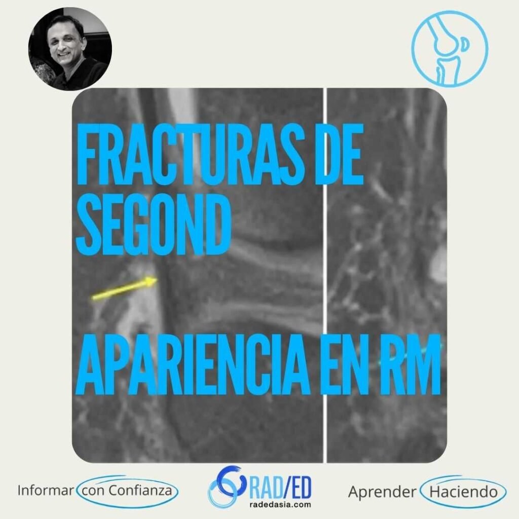

Las fracturas de Segond son fracturas de la meseta tibial lateral que suelen estar asociadas a roturas de LCA. Se cree que están relacionadas con la inserción tibial del ligamento anterolateral. Sin embargo, este ligamento no se identifica con claridad en la RM, y el mejor punto de referencia es observar la zona inmediatamente posterior a la inserción de la banda BIT.

Meseta tibial lateral, justo detrás de la inserción de la banda iliotibial.
Se cree que está relacionado con la inserción tibial del ligamento anterolateral. Sin embargo, este ligamento no se identifica con claridad en la RM, y el mejor punto de referencia es observar la zona inmediatamente posterior a la inserción de la banda BIT.
Pequeña fractura por avulsión o de tipo «escama» de la meseta tibial lateral.
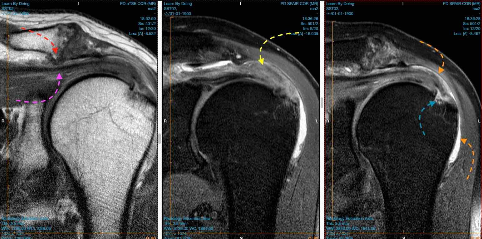

Imagen superior: La radiografía muestra un fragmento de fractura (flecha azul) adyacente a la meseta tibial lateral. El ligamento tibio-tibial (BIT) (flecha amarilla) se encuentra intacto y normal. Tercera imagen de la RM: se observa un edema medular (flecha azul) y una línea de fractura de baja señal inmediatamente posterior a la inserción del BIT.

Imagen superior: Fractura de Segond con inserción normal del BIT (flecha amarilla). Fractura de la cortical lateral (flechas azules) situada detrás de la inserción del BIT.
Una fractura de Segond es una fractura por avulsión de la meseta tibial lateral en el punto de inserción del ligamento longitudinal anterior (LAL). Está estrechamente relacionada con una rotura subyacente del LCA.
Key MRI findings include a a step or break in the black cortical line, bony avulsion fragment at the lateral tibial plateau and in the acute stage bone marrow edema.
Una fractura de Segond suele producirse como consecuencia de una Rotación interna y una tensión en varo de la rodilla. Este mecanismo ejerce una tensión excesiva sobre el ligamento capsular lateral, lo que provoca la avulsión en el punto de inserción del ligamento longitudinal anterior (LAL).