ANKLE

LEARN MSK MRI ANKLE ONLINE

ANKLE MSK MRI RADIOLOGY: We regularly post on Ankle MRI and our Ankle MRI Courses with all the posts on this page.

Ankle MRI can be difficult due to the complex anatomy. In our posts we have concise explanation of the Ankle MRI anatomy, the macroscopic pathology that’s relevant to the Imaging and the Imaging findings in various pathologies.

Learn where to Look, What to Look for and How to Report Ankle MRI More Confidently

mri periosteum callus periosteal new bone radedasia radiology education

PERIOSTEAL REACTION RADIOLOGY MRI: THE RANGE OF TRAUMATIC PERIOSTEAL CHANGES ON MRI

PERIOSTEAL REACTION RADIOLOGY MRI TRAUMATIC PERIOSTITIS & PERIOSTEAL CHANGE WHAT DOES PERIOSTEAL CHANGE SECONDARY TO TRAUMA LOOK LIKE ON MRI There are a number of appearances of periosteal reaction on MRI secondary to trauma that we can see and report. The Histology…Yes Histology!: It helps to have a simple understanding of the histology of the …

PERIOSTEAL REACTION RADIOLOGY MRI: THE RANGE OF TRAUMATIC PERIOSTEAL CHANGES ON MRI Leer más »

mri achilles tendon abnormalities ankle radiology

MRI OF A FEW ACHILLES TENDON ABNORMALITIES: INSERTIONAL TENDONOPATHY TENDONOSIS, BURSA & RUPTURE

ACHILLES TENDON ABNORMALITIES ANKLE MRI ACHILLES TENDON ABNORMALITIES A few MRI images of Achilles pathology from all the findings seen in Achilles Insertional Tendonopathy, Retrocalcaneal Bursitis, Retroachilles Bursitis to MRI of Achilles Rupture. MRI ACHILLES TENDON ABNORMALITIES MRI ACHILLES INSERTIONAL TENDONOPATHY: WHAT TO LOOK FOR MRI findings in Insertional Achilles Tendinopathy: A few findings to …

MRI OF A FEW ACHILLES TENDON ABNORMALITIES: INSERTIONAL TENDONOPATHY TENDONOSIS, BURSA & RUPTURE Leer más »

sesamoid osteonecrosis mri radiology

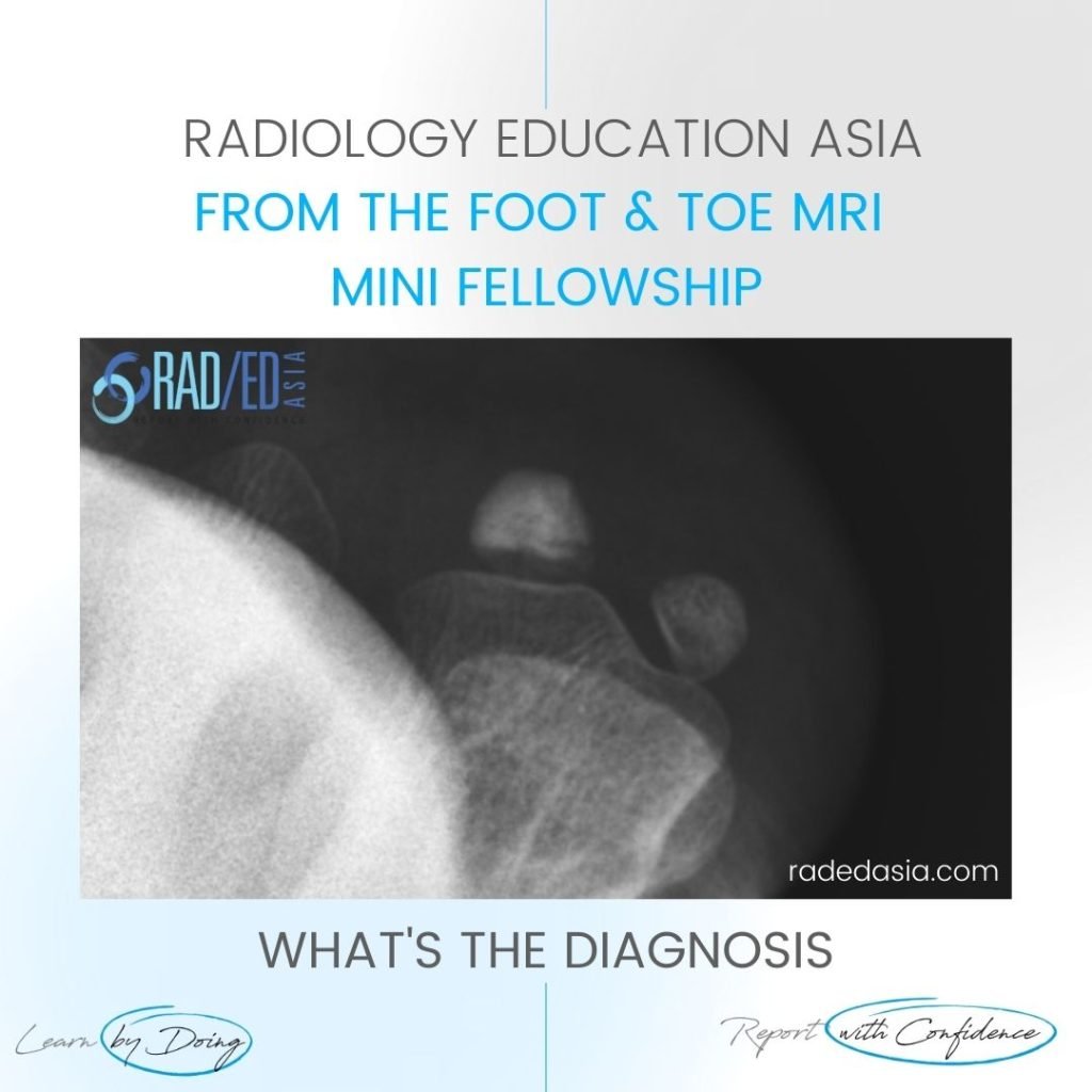
SESAMOID OSTEONECROSIS RADIOLOGY XRAY MRI

SESAMOID OSTEONECROSIS XRAY CT MRI: OVERVIEW OSTEONECROSIS OF THE SESAMOID Osteonecrosis of the sesamoids of the 1st metatarsal is not common but important that it’s recognized. As it progresses fracture, fragmentation and collapse of the sesamoid can occur. SESAMOID OSTEONECROSIS XRAY CT MRI: WHAT TO LOOK FOR SESAMOID OSTEONECROSIS XRAY: WHAT TO LOOK FOR If …

SESAMOID OSTEONECROSIS RADIOLOGY XRAY MRI Leer más »

weight bearing x-rays for lisfranc joint injury instability fracture radedasia

WEIGHT BEARING X-RAYS FOR LISFRANC JOINT INJURY INSTABILITY & FRACTURE (VIDEO)

THE MAIN POINTS TO NOTE OVERVIEW WEIGHT BEARING X-RAYS FOR LISFRANC JOINT INJURY INSTABILITY & FRACTURE   Early diagnosis of Lisfranc instability is crucial to prevent premature OA in the mid foot. Subtle instability patterns can be very difficult to identify on xrays.  Plain radiographs may not effectively identify instability at the LisFranc Joint, especially …

WEIGHT BEARING X-RAYS FOR LISFRANC JOINT INJURY INSTABILITY & FRACTURE (VIDEO) Leer más »

freiberg disease mri radiology toe radedasia

FREIBERG DISEASE SUBCHONDRAL FRACTURE INFRACTION OSTEOCHONDROSIS (VIDEO)

FREIBERG DISEASE / SUBCHONDRAL FRACTURE INFRACTION / OSTEOCHONDROSIS DISCUSSION FREIBERG DISEASE / SUBCHONDRAL FRACTURE INFRACTION / OSTEOCHONDROSIS Extensive oedema in the head of the 2nd metatarsal with slight flattening of the cortical margin is characteristic of what’s called Freiberg’s Disease in adolescents and Subchondral Fracture in adults. Freiberg disease occurs in adolescents with an unfused …

FREIBERG DISEASE SUBCHONDRAL FRACTURE INFRACTION OSTEOCHONDROSIS (VIDEO) Leer más »

ultrasound-ankle-foot-baxter's-nerve-inferior-calcaneal-nerve-entrapment-radiology-radedasia

BAXTER’S NERVE ULTRASOUND HOW TO FIND IT (VIDEO)

BAXTER'S NERVE ULTRASOUND Dr RAJENDRA SAHOO Dr Rajendra Sahoo is a senior consultant Pain Physician and Anesthetist who is Fellowship trained in Ultrasound guided Pain Management. We will be collaborating with Dr Sahoo to bring more MSK and Spine Ultrasound learning. BAXTER’S NERVE ULTRASOUND: HOW TO FIND IT How do you find the Baxter’s nerve …

BAXTER’S NERVE ULTRASOUND HOW TO FIND IT (VIDEO) Leer más »

mri plantar fascia tear fasciitis plantar fascia anatomy heel pain mri mri online course radedasia

MRI PLANTAR FASCIA ANATOMY: WHAT ARE THE 3 BANDS OF THE PLANTAR FASCIA (VIDEO)

MRI PLANTAR FASCIA ANATOMY MRI PLANTAR FASCIA ANATOMY: NORMAL BANDS Today’s post is a video on MRI of the Plantar Fascia Anatomy. We are looking at how to identify the 3 bands of the Plantar Fascia and which band is most important in Plantar Fasciitis. If you find the video helpful, please subscribe to the …

MRI PLANTAR FASCIA ANATOMY: WHAT ARE THE 3 BANDS OF THE PLANTAR FASCIA (VIDEO) Leer más »

peroneal tubercle mri retrotrochlear eminence mri peroneal temdon tondonitis tear radiology education asia(1)

MSK MRI PERONEAL TUBERCLE & RETROTROCHLEAR EMINENCE MRI (VIDEO)

MRI PERONEAL TUBERCLE RETROTROCHLEAR EMINENCE MRI MSK How to identify the Peroneal tubercle and Retrotrochlear eminence on MRI: Today we are looking at MRI of the Peroneal Tubercle and the Retrotrochlear eminence. The importance of these structures is that when enlarged, a Peroneal Tubercle and less commonly the Retrotrochlear eminence, can result in Peroneal Tendonitis, …

MSK MRI PERONEAL TUBERCLE & RETROTROCHLEAR EMINENCE MRI (VIDEO) Leer más »

ankle mri calcaneofibular ligament tear radedasia

CALCANEOFIBULAR LIGAMENT TEAR MRI ANKLE

CALCANEOFIBULAR LIGAMENT TEAR MRI ANKLE CALCANEOFIBULAR LIGAMENT NORMAL MRI The Calcaneofibular ligament is a small lateral ankle ligament that is often torn.   Its normal MRI appearance is a thin low signal structure (Pink arrows) that extends from the calcaneum to the fibula and passes deep to the peroneal tendons (Green arrows).   CALCANEOFIBULAR LIGAMENT …

CALCANEOFIBULAR LIGAMENT TEAR MRI ANKLE Leer más »

ligament retinacula scar mri radiology education asia radedasia

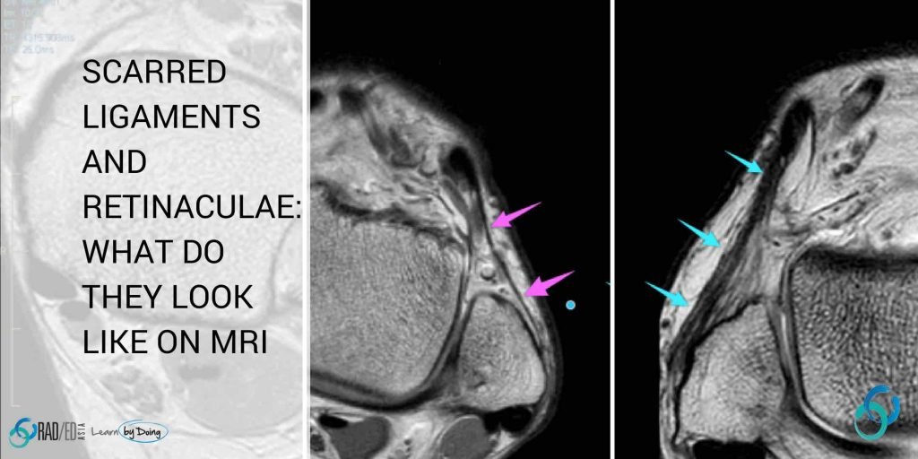
MRI RETINACULA SCAR: SCARRED LIGAMENTS AND RETINACULA: WHAT DOES MRI SCAR TISSUE LOOK LIKE? (VIDEO)

LIGAMENT AND RETINACULA HEALING: WHAT DOES SCAR TISSUE LOOK LIKE ON MRI NEED TO KNOW PATHOLOGY What happens to a ligament when it is torn and injured? This is a brief edited summary from an article on the pathology of scar formation ( “Scar formation and ligament healing” link below).   “After an injury… hemostasis …

MRI RETINACULA SCAR: SCARRED LIGAMENTS AND RETINACULA: WHAT DOES MRI SCAR TISSUE LOOK LIKE? (VIDEO) Leer más »

Este sitio está destinado únicamente a profesionales médicos. El uso de este sitio se rige por nuestra Términos de servicio y Declaracion de privacidad que se puede encontrar haciendo clic en los enlaces. Acepte antes de continuar con el sitio web.
error: Clic derecho no disponible.