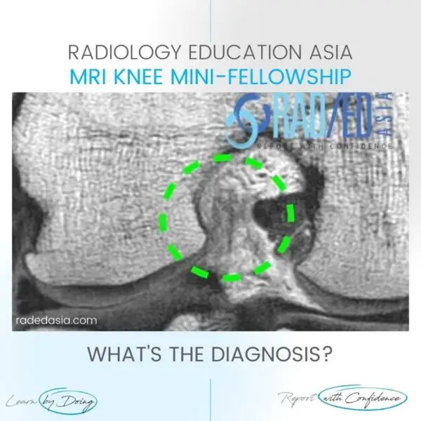
- This is a facet synovial cyst.
- These arise from the facet joint and can expand in any direction.
- Look for a well defined , walled structure that follows fluid signal on all sequences.
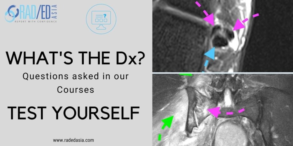
Well defined cystic structure (Pink arrow) adjacent to the facet joint and extending into the canal in the epidural space.
![facet synovial cyst radiology lumbar spine mri facet synovial cyst radiology lumbar spine mri](https://radedasia.com/wp-content/uploads/2022/02/facet-synovial-cyst-radiology-lumbar-spine-mri.jpg)
![facet synovial cyst radiology mri lumbar spine facet synovial cyst radiology mri lumbar spine](https://radedasia.com/wp-content/uploads/2022/02/facet-synovial-cyst-radiology-mri-lumbar-spine.jpg)
#radiology #radedasia #mri #spinemri #mskmri #mrispine #radiologyeducation #radiologycases #radiologist #radiologycme #radiologycpd #medicalimaging #imaging #radcme #spinedegeneration #rheumatology #arthritis #rheumatologist #degenerativedisease #orthopaedic #painphysician #chiropractic #chiropracter #physiotherapy #sportsmed #orthopaedic #mskmri #facetcyst
#radedasia #mri #mskmri #radiología