MENISCOTIBIAL LIGAMENT TEAR DEEP MCL MRI KNEE
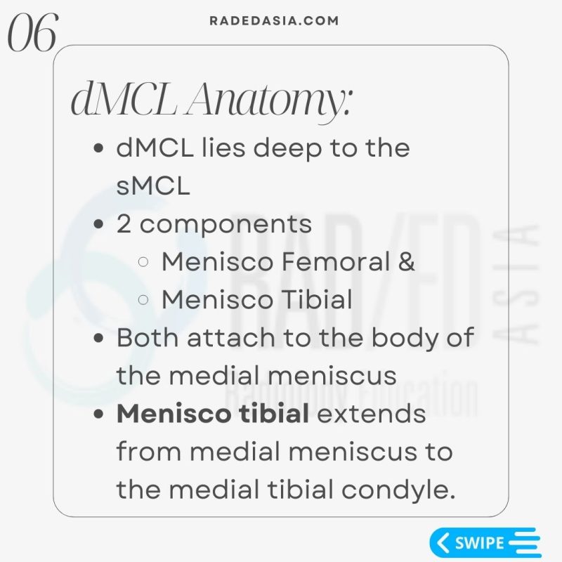
- The dMCL lies deep to the sMCL.
- It has 2 components.
- Menisco Femoral &,
- Menisco Tibial.
- Both attach to the body of the medial meniscus
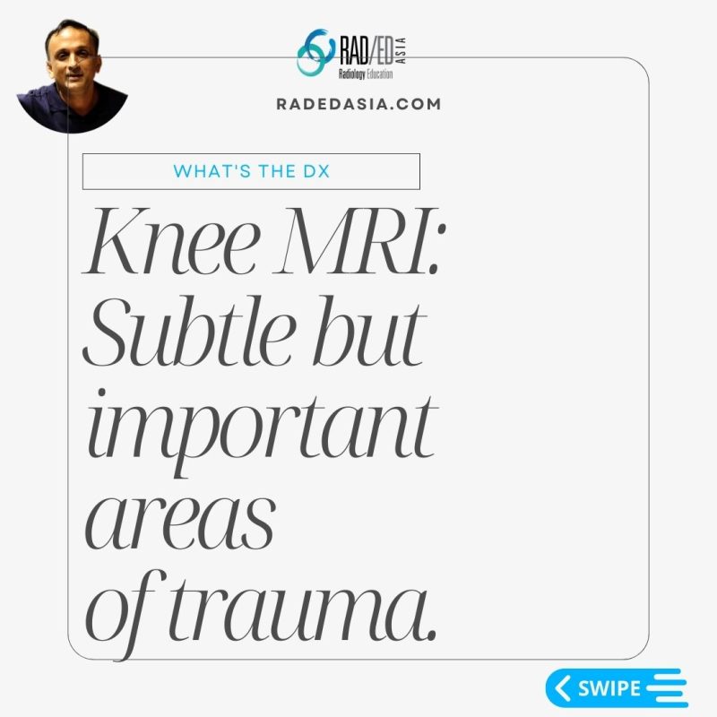
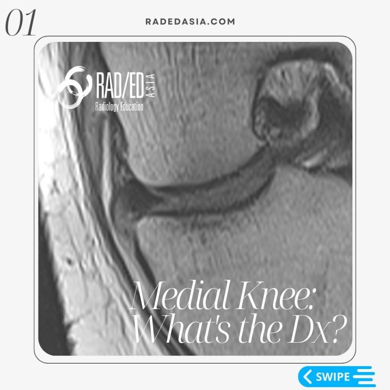
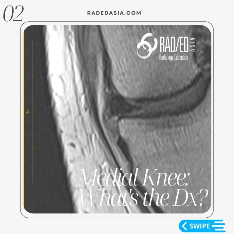
- The Meniscotibial ligament extends from medial meniscus to the medial tibial condyle.

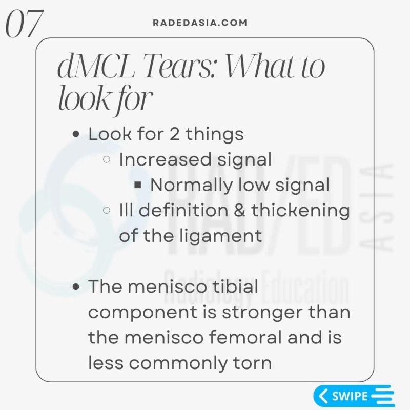
- Look for 2 things,
- Increased signal of the ligament.
- Normally the menisco-tibial ligament is low signal.
- Ill definition & thickening of the ligament.
- Increased signal of the ligament.
- The menisco-tibial component is stronger than the menisco-femoral and is less commonly torn.

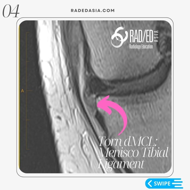
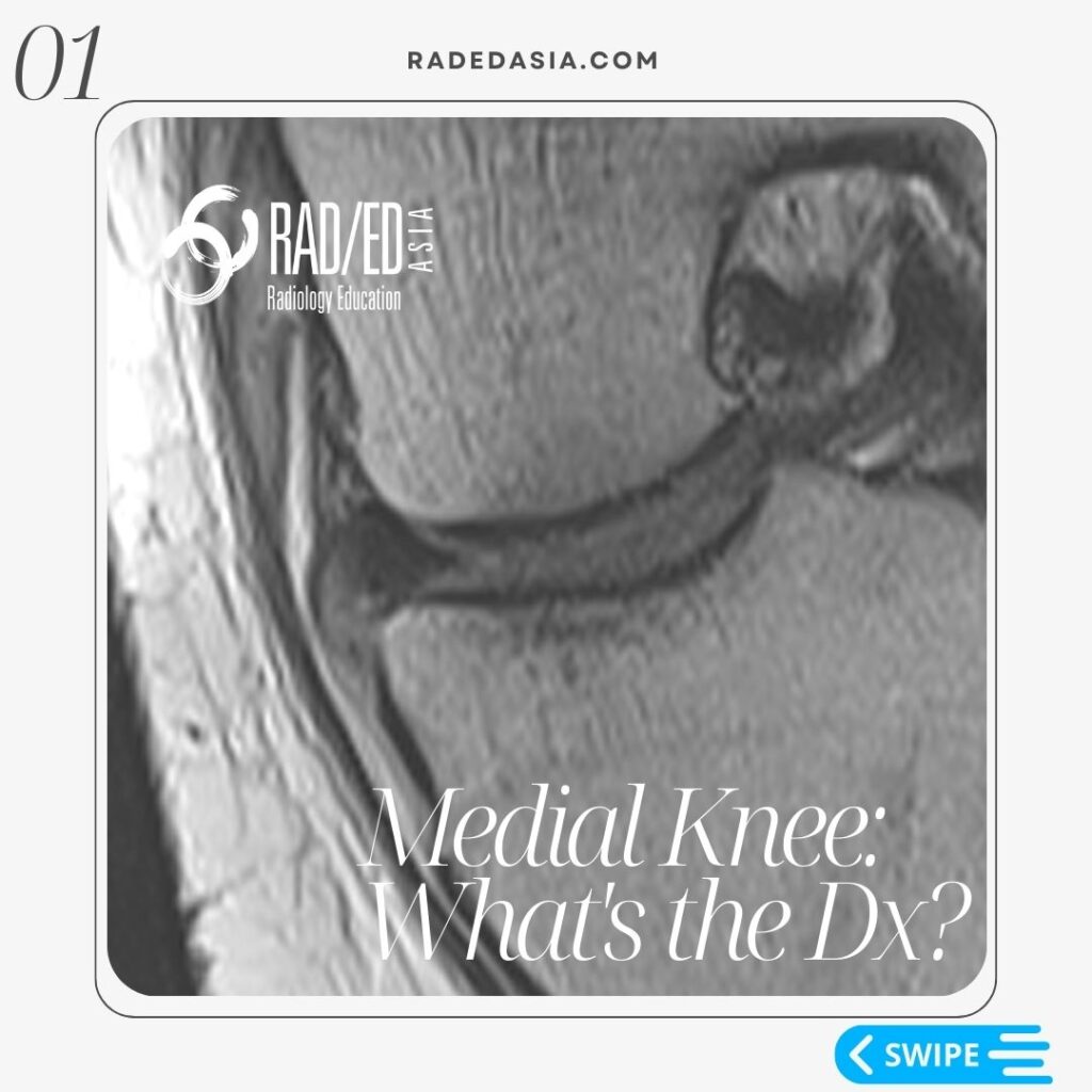
In this case the menisco-tibial ligament is hyper-intense, thickened and ill defined (Pink arrows). All features of a torn menisco-tibial ligament.
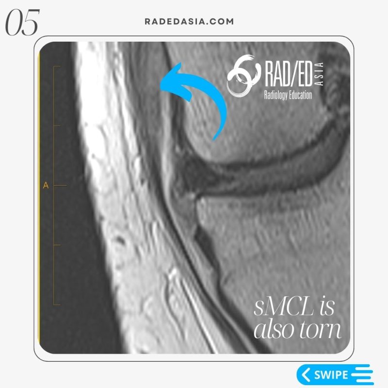
You can also see that similar changes are present in the proximal sMCL which is also torn (Blue arrow).

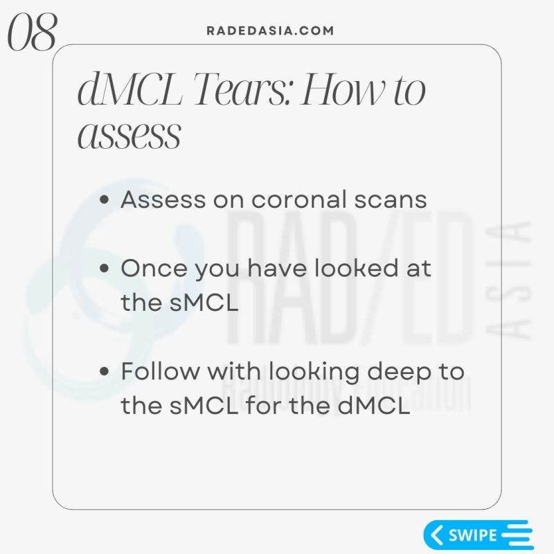
- Assess on coronal scans the menisco-tibial and menisco-femoral ligaments.
- Start by assessing the sMCL.
- Once you have looked at the sMCL look immediately deep to it to find the deep MCL components.


