Anterior Cruciate Ligament

acl-pcl-mucoid-degeneration-mri-findings-radedasia

ACL & PCL MUCOID DEGENERATION MRI FINDINGS

MRI FINDINGS IN ACL AND PCL MUCOID DEGENERATION Mucoid degeneration can affect both the anterior cruciate ligament (ACL) and posterior cruciate ligament (PCL). Mucoid degeneration is often confused with ACL tears and this post looks at the main MRI features of ACL and PCL mucoid degeneration that will help to differentiate from tears. MRI FINDINGS …


ACL & PCL MUCOID DEGENERATION MRI FINDINGS Leer más »

acl tear mri segond fracture radiology education asia online radiology mri course

SEGOND FRACTURE MRI: WHERE TO LOOK, WHAT TO LOOK FOR, HOW TO REPORT (VIDEO)

MRI SEGOND FRACTURE KNEE ACL TEAR Segond Fracture MRI , How to find it when Assessing the ACL on MRI & what ACL associated injuries to look for: Today’s video post is on MRI of Segond Fracture which is one of the injuries associated with ACL tears. We look at where to look, what to …

SEGOND FRACTURE MRI: WHERE TO LOOK, WHAT TO LOOK FOR, HOW TO REPORT (VIDEO) Leer más »

subcortical subchondral fracture knee tibial plateau mri insufficiency

SUBCORTICAL SUBCHONDRAL FRACTURE KNEE MRI TIBIAL PLATEAU (VIDEO)

SUBCORTICAL SUBCHONDRAL FRACTURE KNEE MRI TIBIAL PLATEAU DISCUSSION Subchondral or subcortical fractures of the knee can occur in the femoral condyles or tibial plateau.   The key to the diagnosis is seeing a linear low signal line adjacent to and paralleling the cortex without any cortical defect or break in the acute phase.     There …

SUBCORTICAL SUBCHONDRAL FRACTURE KNEE MRI TIBIAL PLATEAU (VIDEO) Leer más »

mri acl tear pivot shift injury bone contusion pattern osteochondral fracture knee

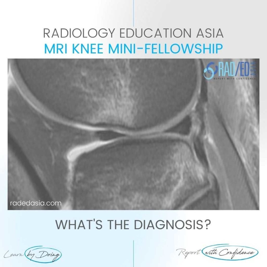
ACL TEAR PIVOT SHIFT INJURY BONE CONTUSION PATTERN OSTEOCHONDRAL FRACTURE MRI KNEE

ACL TEAR PIVOT SHIFT INJURY BONE CONTUSION PATTERN OSTEOCHONDRAL FRACTURE MRI WHAT'S THE Dx? Pivot shift bone bruising of the lateral femoral condyle and an osteochondral depression fracture secondary to an ACL tear.   Characteristic bone contusion pattern seen with complete ACL tear.   The osteochondral fracture is seen with the flattening of the cortical …

ACL TEAR PIVOT SHIFT INJURY BONE CONTUSION PATTERN OSTEOCHONDRAL FRACTURE MRI KNEE Leer más »

medullary infarct radiology femur tibia mri radedasia

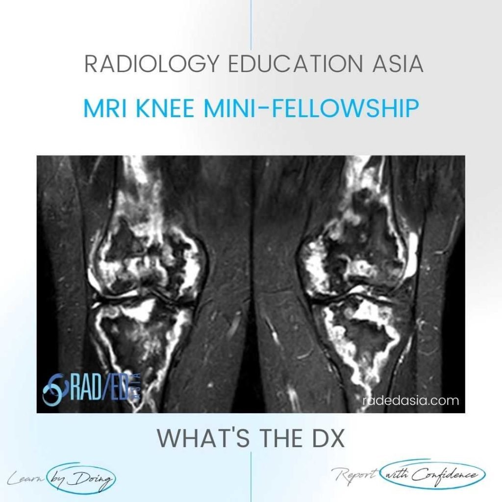
MEDULLARY BONE INFARCTS RADIOLOGY KNEE MRI

MOVE SLIDER ICON (ARROWS) LEFT TO SEE THE DIAGNOSIS FINDINGS Bilateral medullary femoral and tibial serpiginous STIR hyper-intensity with extension into the femoral and tibial epiphysis. DIAGNOSIS MEDULLARY BONE INFARCTS. This is a characteristic appearance of medullary bone infarcts. Most commonly they are in the diaphysis and metaphysis but in this case they also extend …

MEDULLARY BONE INFARCTS RADIOLOGY KNEE MRI Leer más »

arcuate segond fracture fibular styloid radiology education asia tuesday tip

ARCUATE FRACTURE KNEE X-RAY, SEGOND FRACTURE, LATERAL KNEE FRACTURES

ARCUATE FRACTURE, SEGOND FRACTURE, LATERAL KNEE FRACTURES ARCUATE FRACTURE KNEE X-RAY , SEGOND FRACTURE, LATERAL FIBULAR FRACTURE This post looks at the 3 fractures (Segond Fracture, Arcuate Fracture and Lateral Fibular Fracture ) that can be seen on the lateral aspect of the knee x-ray which are indicative of underlying tendon or ligament involvement. The …

ARCUATE FRACTURE KNEE X-RAY, SEGOND FRACTURE, LATERAL KNEE FRACTURES Leer más »

Este sitio está destinado únicamente a profesionales médicos. El uso de este sitio se rige por nuestra Términos de servicio y Declaracion de privacidad que se puede encontrar haciendo clic en los enlaces. Acepte antes de continuar con el sitio web.
error: Clic derecho no disponible.