Rótula

mri knee fat pad impingement radiology education asia

KNEE MRI FAT PAD IMPINGEMENT: WHERE TO LOOK & WHAT TO LOOK FOR

KNEE FAT PAD IMPINGEMENT MRI APPEARANCE MRI OF FAT PAD IMPINGEMENT AROUND THE KNEE   Knee fat pad impingement syndromes are characterized by pain and inflammation of the fat pads surrounding the knee joint and can significantly impact knee function. This blog post looks at the anatomy of the knee fat pads, the MRI appearance …

KNEE MRI FAT PAD IMPINGEMENT: WHERE TO LOOK & WHAT TO LOOK FOR Leer más »

knee mri bursa suprapatellar plica

KNEE MRI PLICA SUPRAPATELLAR BURSA LIPOHEMARTHROSIS

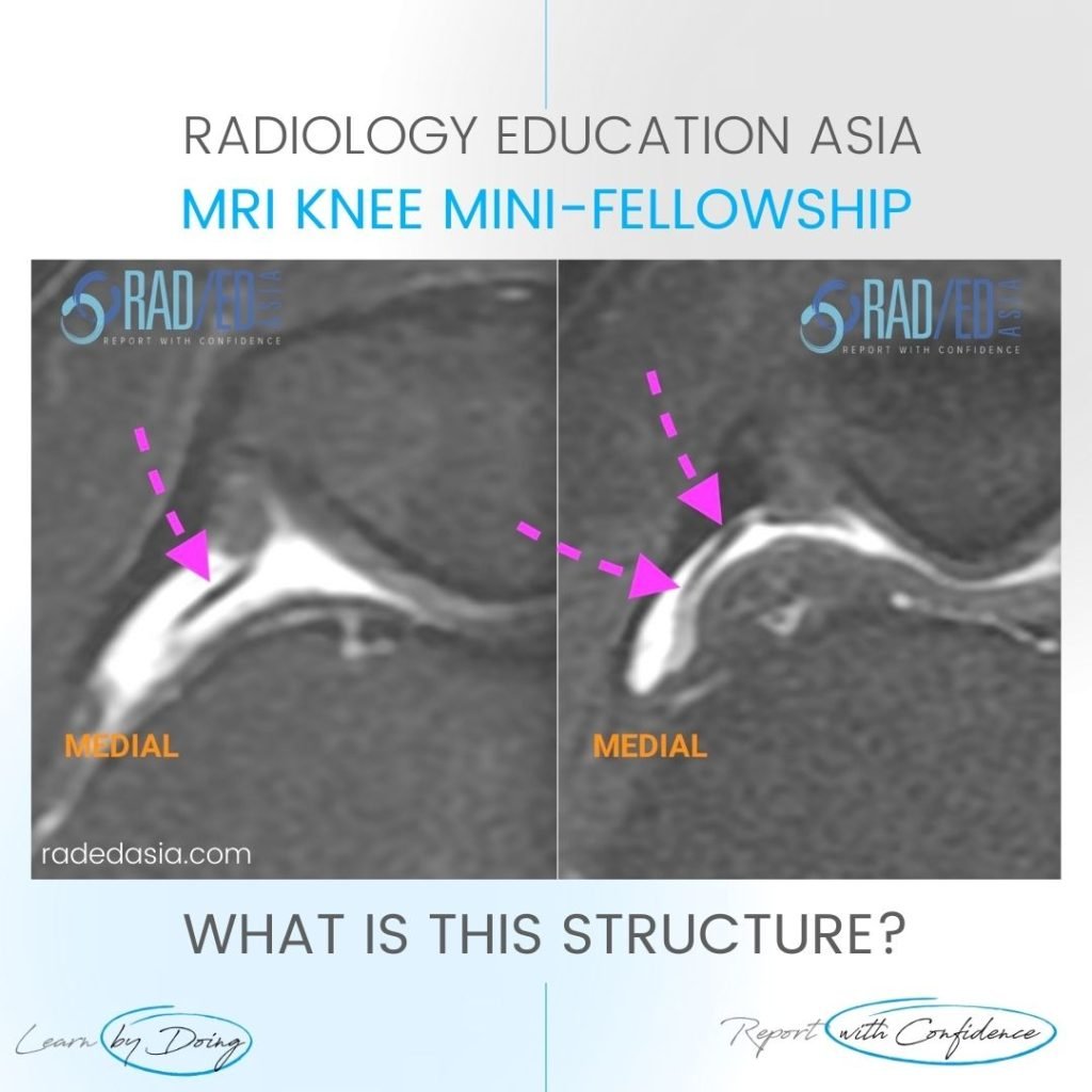
KNEE MRI PLICA SUPRAPATELLAR BURSA LIPOHEMARTHROSIS NORMAL SUPRA PATELLAR PLICA The normal supra patellar plica is not continuous.  There is a gap in it ( fenestration) which allows fluid to circulate freely in the SP bursa above and below the plica. CONTINUOUS SUPRAPATELLAR PLICA At times the SP Plica is continuous with no gap.  Normally …

KNEE MRI PLICA SUPRAPATELLAR BURSA LIPOHEMARTHROSIS Leer más »

learn mri knee mucoid degen radedasia

ACL MUCOID DEGENERATION RADIOLOGY MRI

ACL MUCOID DEGENERATION RADIOLOGY MRI DISCUSSION Mucoid Degeneration. These findings indicate Mucoid Degeneration of the ACL rather than a tear.   This is a characteristic appearance of ACL mucoid degeneration where the ACL is expanded and hyperintense but is still continuous with normal alignment. . WHAT ARE THE FINDINGS The ACL is enlarged and hyperintense …

ACL MUCOID DEGENERATION RADIOLOGY MRI Leer más »

patella dislocation subluxation fracture hemarthrosis lipohemarthrosis radiology mri xray knee

PATELLA DISLOCATION SUBLUXATION FRACTURE HEMARTHROSIS LIPOHEMARTHROSIS MRI KNEE RADIOLOGY

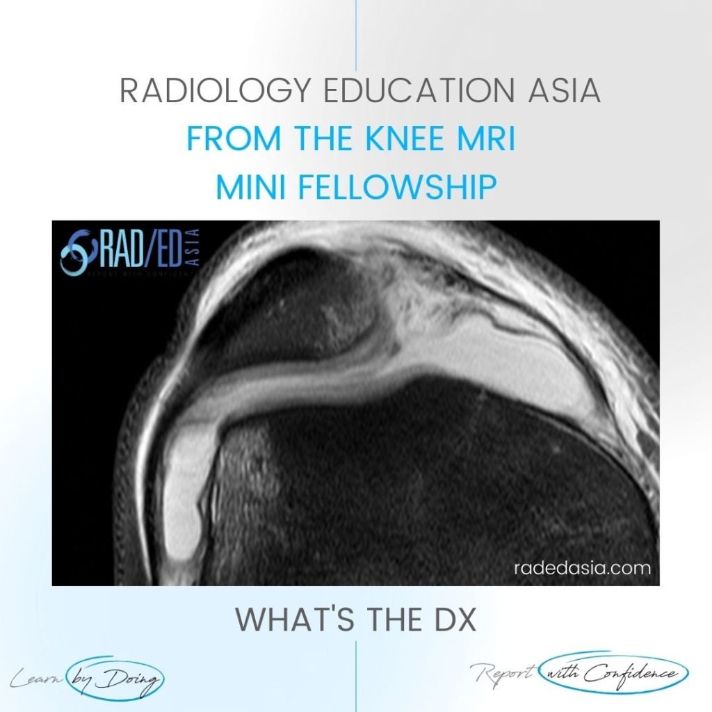
PATELLA DISLOCATION SUBLUXATION HEMARTHROSIS FRACTURE DISCUSSION ON PATELLA DISLOCATION SUBLUXATION HEMARTHROSIS FRACTURE The patient has had a recent patella dislocation and there is now a large suprapatellar effusion (on MRI this was a hemarthrosis).  On X-ray we can’t say defnitely that it’s a hemarthrosis but the presence of a fracture would indicate that it is …

PATELLA DISLOCATION SUBLUXATION FRACTURE HEMARTHROSIS LIPOHEMARTHROSIS MRI KNEE RADIOLOGY Leer más »

meniscus cyst radiology intrameniscal tear mri knee

MENISCAL CYST RADIOLOGY INTRAMENISCAL TEAR MRI KNEE

MENISCAL INTRAMENISCAL CYST MRI RADIOLOGY DISCUSSION INTRAMENISCAL CYST. Extensive intra meniscal cyst anterior horn. A tear is not seen extending to the articular margin. However we may not always see the extension on MRI. So if you see an intra-meniscal or parameniscal cyst these are always due to an underlying tear even if we don’t …

MENISCAL CYST RADIOLOGY INTRAMENISCAL TEAR MRI KNEE Leer más »

patellar tendon trauma contusion mri

PATELLAR TENDON CONTUSION TRAUMA MRI

PATELLAR TENDON CONTUSION Patellar Tendon Contusion. In this case we have Patellar tendon contusion and Patella contusion.   Most commonly this combined appearance would be of Patellar Tendonosis with reactive oedema in the patella and inflammatory change extending into Hoffa’s fat.   However the history of trauma in this patient is important in being able …

PATELLAR TENDON CONTUSION TRAUMA MRI Leer más »

mri knee course patella dislocation

KNEE MRI PATELLA DISLOCATION SUBLUXATION

KNEE MRI PATELLA DISLOCATION SUBLUXATION DISCUSSION PATELLA DISLOCATION Patella Dislocation Subluxation MRI.  There has been a recent Patella dislocation with classical contra coup bone bruising and torn MPFL/ medial retinaculum.   The best clue to patella dislocation / subluxation on MRI is the presence of the contra coup bone bruising in the patella and femur. …

KNEE MRI PATELLA DISLOCATION SUBLUXATION Leer más »

patellar dislocation lateralisation tibial tubercle radiology education asia

MRI TIBIAL TUBERCLE HOW TO ASSESS LATERALISATION OF THE TIBIAL TUBEROSITY

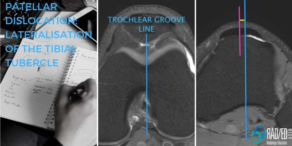
MRI TIBIAL TUBERCLE MRI TIBIAL TUBERCLE : HOW TO ASSESS LATERALISATION OF THE TIBIAL TUBERCLE   Lateralization of the tibial tubercle is one of the predisposing factors for Patellar Dislocation.    In this post we look at what the normal position is, how to measure lateralization and what the differences between CT and MRI measurements …

MRI TIBIAL TUBERCLE HOW TO ASSESS LATERALISATION OF THE TIBIAL TUBEROSITY Leer más »

Este sitio está destinado únicamente a profesionales médicos. El uso de este sitio se rige por nuestra Términos de servicio y Declaracion de privacidad que se puede encontrar haciendo clic en los enlaces. Acepte antes de continuar con el sitio web.
error: Clic derecho no disponible.