
OVERVIEW:
Meniscal Ossicles are the areas of ossification within the meniscus. In this post we look at What Are They, Where to Look and How to Report well.
Area of ossification within the meniscus.
This is a nice article from AJR on Meniscal Ossicles which indicates that they are post traumatic in origin. (Meniscal Ossicle: Post-traumatic Origin and Association with Posterior Meniscal Root Tears. Mohankumar et al AJR 2014; 203:1040-1046).
Most common location was in the meniscal root or posterior horn of the medial meniscus. Much more uncommon in the lateral meniscus.

(Table from Reference above)
Two possible causes:
1. Post traumatic metaplasia and heterotopic ossification of a meniscal tear (either posterior horn or root).
2. Avulsion of bone related to a meniscal root tear (less common).
Ossicles are iso-intense to bone marrow on all sequences.
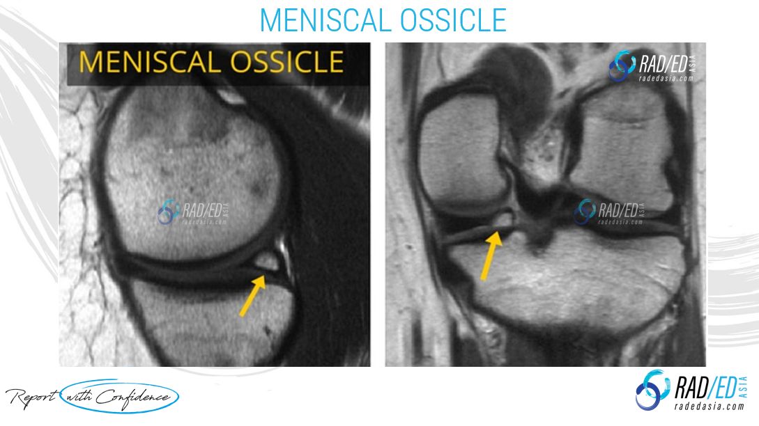
Image above: well corticated meniscal ossicle (yellow arrow) following bone marrow signal. (Image Radiology Education Asia)
Be Careful of this:
Look at both the PD and PDFS/ T2FS sequences together as you may miss it if you look only at the PDFS/ T2FS sequence. This is because it contains marrow fat which will loose signal on PDFS and make the ossicle look like normal meniscus.
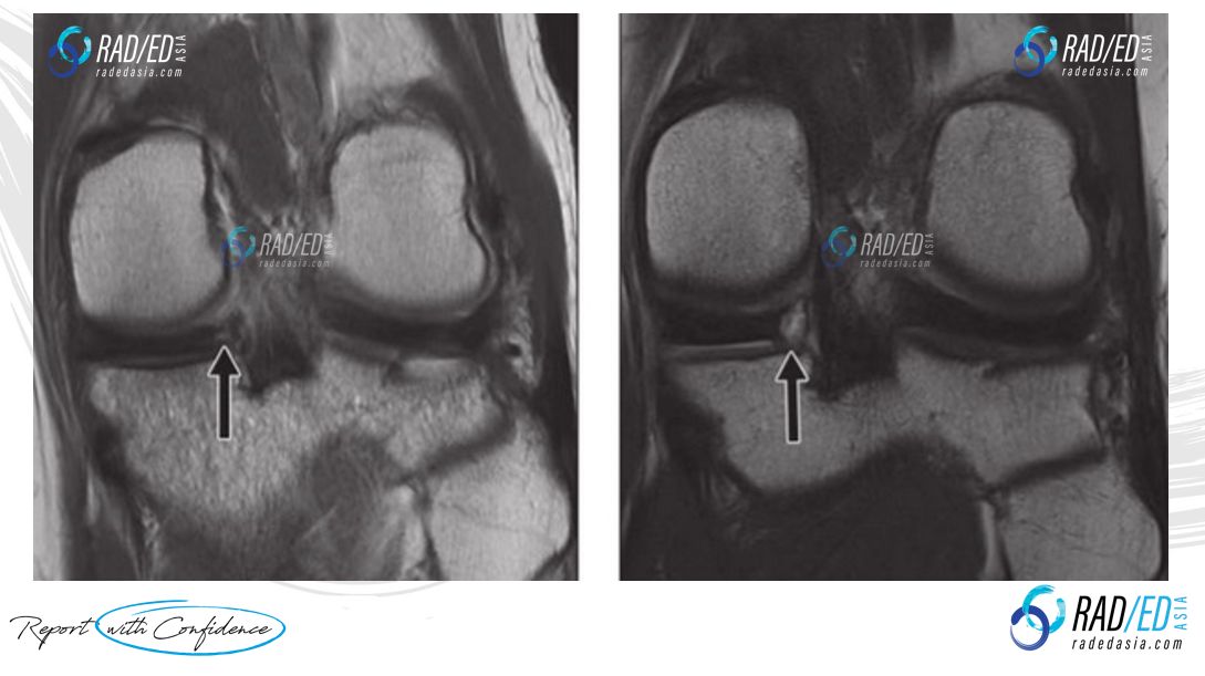
Two images below demonstrate interval development of Ossicles and temporal increase in size.

Image Above: Image A initial scan normal posterior horn/ root. Image B 33 months later development of meniscal ossicle (arrow). Images from credited article.