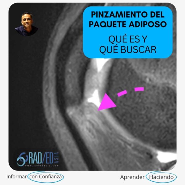
POSTERIOR HORN MENISCUS: Three posts for such a small thing. How can it be so important?
ANATOMY: The posterior horn has two components
1. The posterior horn itself
2. The meniscal root which attaches the posterior horn to the intercondylar fossa
TYPES OF TEARS: The posterior horn can have the usual types of meniscus tears (horizontal cleavage, vertical, radial, complex) but there are two specific types of tears that are easy to miss.
THE AXE MURDERER TEAR: Ok I made that name up and if you are a trainee dont use the term in your exams, but to me it looks like someone has taken an axe to the meniscus and chopped away a portion of the posterior horn usually near the intercondylar fossa.
 The above images are anterior to posterior and in Image B (blue arrow) shows the normal posterior horn of the lateral meniscus extending into the intercondylar fossa. However the posterior horn of the medial meniscus is torn and truncated like its been cut with an axe ( Image C yellow arrow) and you never see it extending to the intercondylar fossa.
Its very easy to miss this on the sagittal scans because the meniscus ends and we don’t notice that it should be extending into the intercondylar fossa. Our eyes and brain are conditioned to look for the presence, and not the absence of something. Its best seen on the coronal images and its important when we assess the meniscus that we look at the posterior horn carefully on the coronal images to make sure that the posterior horns extend into the intercondylar fossa and dont terminate abruptly.
 Truncated medial meniscus posterior horn and meniscal root ( yellow arrows). Lateral meniscus posterior horn and root extend into the intercondylar fossa.
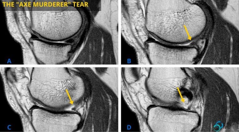
Sagittal scans. Posterior horn and root progressively become smaller. No meniscus in the intercondylar fossa in image D. The absence of meniscus which is the tear is less obvious than on the coronal scans.
MACERATION: Maceration is not a focal tear but its when the meniscus has been ground down and what you see is a remnant that has the rough shape of the meniscus but there is no normal meniscus. Its a ghost like remnant. Its not specific to the posterior horn but is often seen there.
 In images A and B there is maceration with the suggestion of suggestion of a meniscus but its not normal. The shape is roughly retained but the meniscus is abnormal. Coronal image C demonstrates grey material that is the shape of the meniscus but is clearly abnormal.
 Maceration: Image above form of the meniscus is present but content is very abnormal with increased PD signal.
Part 2 next week looks at the the Meniscal Root and tears of the meniscal root.
