
mri ct osteitis condensans ilii spine sij

OSTEITIS CONDENSANS ILII RADIOLOGY CT SACROILIAC JOINT SIJ (VIDEO)

DISCUSSION ON OCI

Osteitis Condensans Ilii.

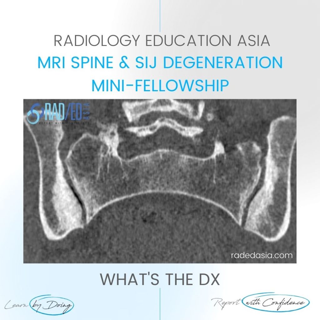
Typical CT sclerotic changes of Osteitis Condensans Ilii. The sclerosis is continuous with the SIJ margin and particularly on the patient’s right it has a triangular appearance.

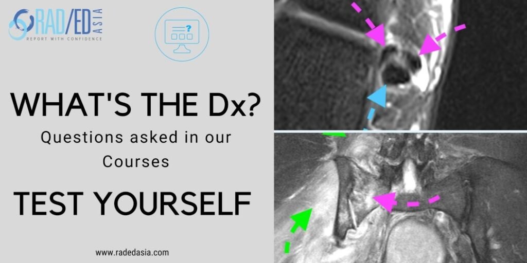
There is also mild sclerosis in the right sacrum adjacent to the SIJ (Green arrow). Sclerosis of the sacrum can also be seen in OCI but it’s not as prominent as the ilium and doesn’t occur in isolation.

Important negative findings are preservation of the SIJ joint space and no erosions.

oci osteitis condensans ilii spine sij ct mri radedasia


Image Above:
Bilateral iliac sclerosis (Pink arrows) adjacent to the SIJ.

Our CPD & Learning Partners

Learn more about SPINE Imaging in our ONLINE Guided SPINE & SIJ Imaging Mini-Fellowship.


More by clicking on the images below.

RM de aracnoiditis que demuestra los patrones normales de realce poscontraste de las raíces nerviosas espinales y las meninges para una interpretación precisa de las imágenes.
Cordón
MRI arachnoiditis illustrating pathogenesis and normal cauda equina nerve root distribution.
Cordón
error: Content is protected !!

Suscríbase a nuestro boletín

Esté atento a los nuevos lanzamientos y aprendizajes de las Fellowships

Fellowships

Mejor que las conferencias

Únase a nuestra comunidad de WhatsApp

Ayudamos a los radiólogos a aprender
haciendo e informa con confianza

Este sitio está destinado exclusivamente a las profesiones médicas. El uso de este sitio se rige por nuestras Condiciones de servicio y Declaración de privacidad, que puede consultar haciendo clic en los enlaces. Por favor, acéptelas antes de continuar en el sitio web.