PAIN MANAGEMENT
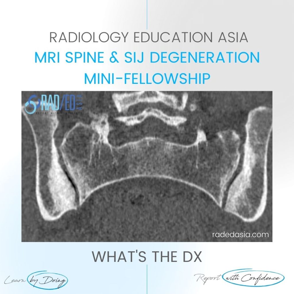
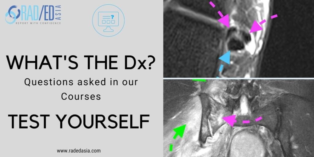
Osteitis Condensans Ilii.
Typical CT sclerotic changes of Osteitis Condensans Ilii. The sclerosis is continuous with the SIJ margin and particularly on the patient’s right it has a triangular appearance.
There is also mild sclerosis in the right sacrum adjacent to the SIJ (Green arrow). Sclerosis of the sacrum can also be seen in OCI but it’s not as prominent as the ilium and doesn’t occur in isolation.
Important negative findings are preservation of the SIJ joint space and no erosions.

Image Above: Bilateral iliac sclerosis (Pink arrows) adjacent to the SIJ.
![]()