MRI of the lumbar spine showing an acute Schmorl's node with characteristic vertebral body changes.

Stay tuned on new Fellowships and learnings

SCHMORL’S NODE ON SPINE MRI: ACUTE & CHRONIC SCHMORL’S NODES MRI

MRI ACUTE AND CHRONIC SCHMORL'S NODES

This post highlights the key imaging appearances of acute and chronic Schmorl’s nodes on spine MRI.

radiology-education-asia-radedasia
 

SCHMORL'S NODES ON SPINE MRI

MRI image of acute Schmorl’s node in the spine showing peripheral bone marrow edema.
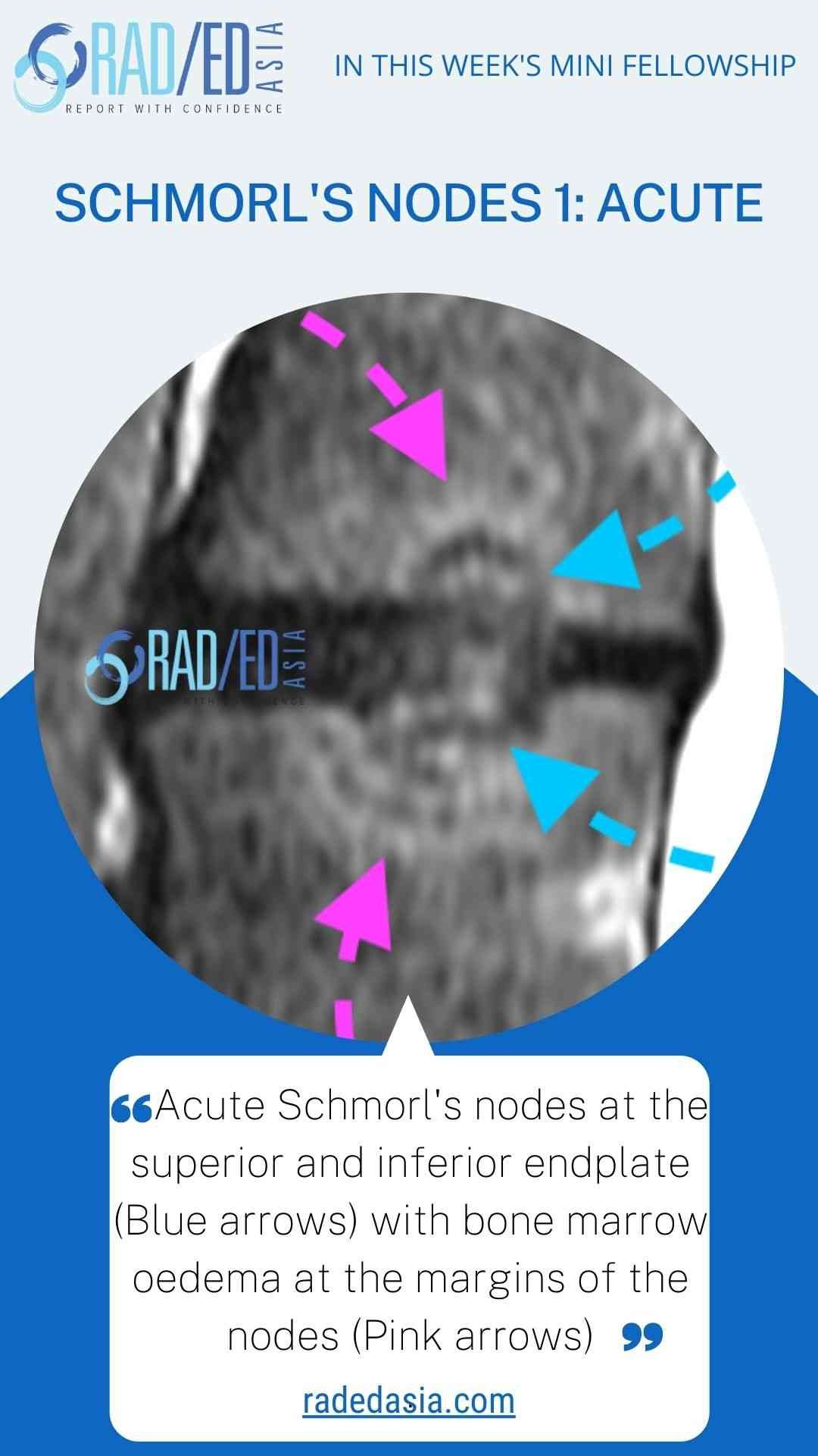
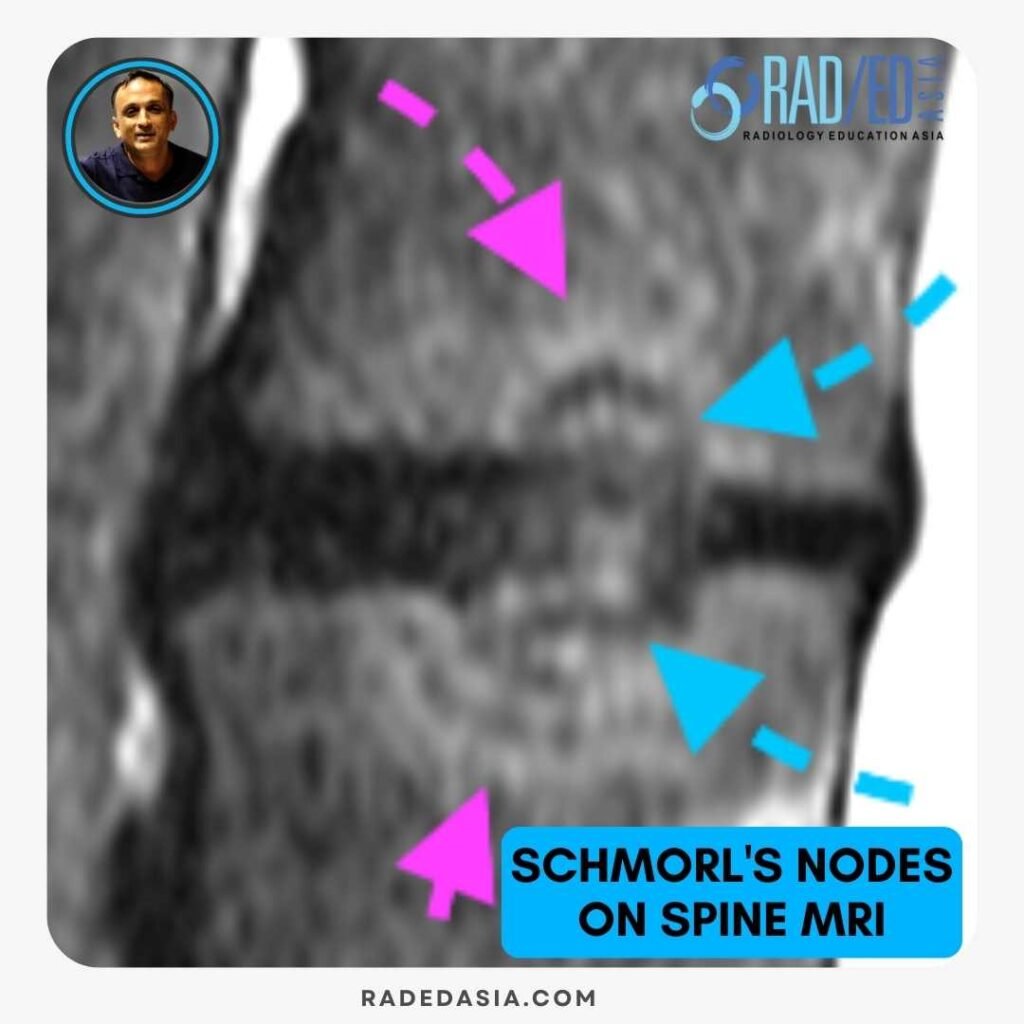
Acute Schmorl’s nodes demonstrate oedema at their margins. This is usually limited to around the circumference of the margins but occasionally can be very extensive.
radiology-education-asia-radedasia

Chronic Schmorl’s node on spine MRI with corticated margins and no edema or fat infiltration.
Chronic Schmorl’s nodes can have a variable appearance. Here the margin is corticated and the marrow signal at the margins of the schmorl’s node is normal with no oedema or fatty change.
radiology-education-asia-radedasia

MRI showing chronic Schmorl’s node in the spine with reactive fatty marrow infiltration.
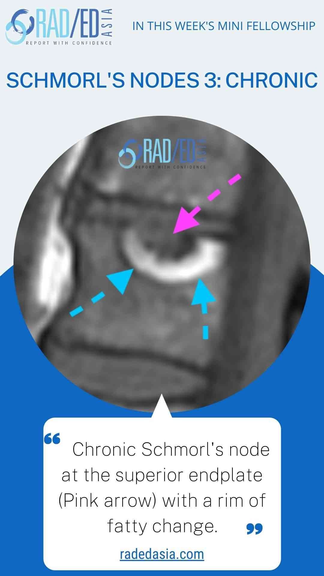
The second appearance of chronic Schmorl’s nodes is for the marrow around the margin to have fatty infiltration, with fat signal more than in the adjacent marrow.
radiology-education-asia-radedasia

Read Article: “Differentiation of Schmorl’s Nodes from Bone Metastases of the Spine, Read HERE

radiology-education-asia-radedasia

TEST YOURSELF ON SOME COMMON & FREQUENTLY ASKED QUESTIONS

Acute Schmorl’s nodes typically show oedema at their margins. This oedema is usually confined around the circumference of the node, though in some cases, it may extend more extensively into the surrounding marrow.
radiology-education-asia-radedasia

Chronic Schmorl’s nodes can present in two forms: some have corticated margins with normal marrow signal and no associated oedema or fatty change; others show fatty infiltration at the node’s margins, which appears as increased fat signal compared to the adjacent marrow.
radiology-education-asia-radedasia

The signal changes—whether oedema in acute cases or fatty infiltration in chronic ones—represent reactive bone marrow changes. These occur in response to disc material herniating into the vertebral body, which triggers inflammation or fat replacement depending on the chronicity.
radiology-education-asia-radedasia

We look at all of these topics in more detail in our Guided SPINE & SIJ Imaging Mini Fellowships.

Click on the image below for more information.

Our CPD & Learning Partners

OTHER POPULAR SPINE REPORTING POSTS: CLICK ON THE IMAGES BELOW

PAIN MANAGEMENT
guillain-barre-syndrome-mri-findings-nerve-enhancement-spine-radiology-radedasia
SPINE DEGENERATION

Stay tuned on new Fellowships and learnings

error: Content is protected !!

Join our Newsletter

Stay tuned on new
Mini-Fellowships launches and learnings

Mini-Fellowships

Better Than Conferences

Join our WhatsApp Community

We help radiologists learn
by doing and report with confidence

This site is intended for Medical Professions only. Use of this site is governed by our Terms of Service and Privacy Statement which can be found by clicking on the links. Please accept before proceeding to the website.