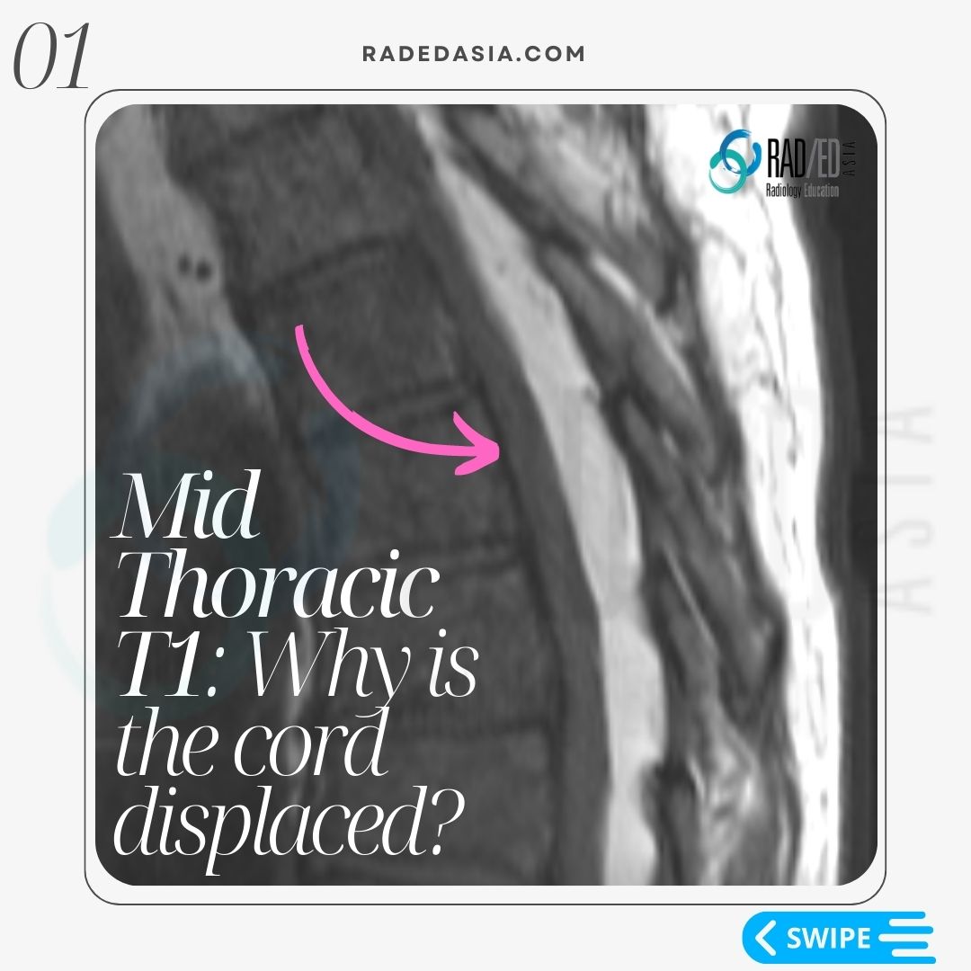

OVERVIEW
Thoracic Epidural lipomatosis is not uncommon to see but has a different appearance to Lumbar Epidural Lipomatosis
![]()
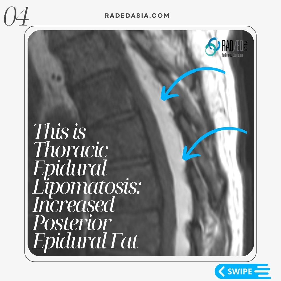
WHAT IS THORACIC EPIDURAL LIPOMATOSIS?
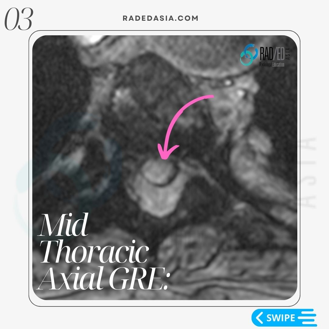
With thoracic epidural lipomatosis there is excess fat accumulated in the epidural space.
WHAT ARE THE CAUSES OF EPIDURAL LIPOMATOSIS?
There is an increased risk of developing epidural lipomatosis with:
- Chronic corticosteroid use.
- Cushing syndrome.
- Obesity.
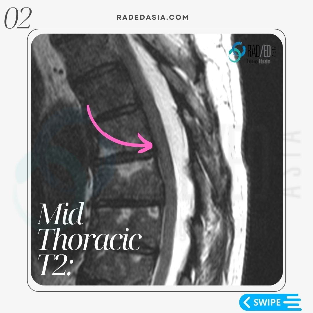
WHAT TO LOOK FOR: MRI OF THORACIC EPIDURAL LIPOMATOSIS
- Epidural lipomatosis in the thoracic spine has a different appearance to Lumbar Epidural Lipomatosis.
- In the lumbar spine we look for a trefoil appearance of the thecal sac.
- However, in the thoracic spine we look for Increased epidural fat.
- The fat accumulation is posterior and lateral.
- This results in the Cord being displaced anteriorly and thecal sac effaced.
- There is no trefoil appearance like you see in the lumbar spine.
![]()
IS THERE A MEASUREMENT?
Posterior epidural fat measuring >6mm in AP diameter is abnormal.
THE TAKE AWAY
- Epidural lipomatosis in the thoracic spine has a different appearance to Lumbar Epidural lipomatosis (See Here).
- Look for increased fat in the epidural space and cord displacement.
![]()