
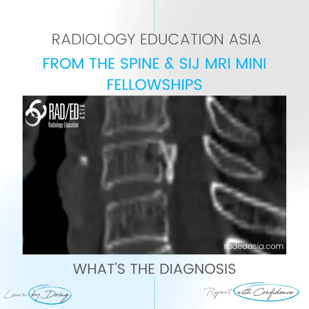
opll spine ossification posterior longitudinal ligament ct mri xray radiology (1)

OPLL SPINE OSSIFICATION POSTERIOR LONGITUDINAL LIGAMENT CT (VIDEO)

  • There is localised, linear ossification present at the posterior margin of the C6 vertebra.
  • This has a typical appearance of OPLL (Ossification of the Posterior Longitudinal Ligament).
  • The cervical spine is the most common location.
  • It is important to follow this up with an MRI of the cord to assess for cord compression and myelopathy.

Ossification present in the midline at the posterior body margin (Pink arrows).

If your Browser is blocking the video, Please view it on our YouTube Channel HERE

Learn more about SPINE Imaging in our ONLINE
Guided MRI Degenerative SPINE Mini-Fellowship.


More by clicking on the images below.

MRI arachnoiditis demonstrating normal post-contrast enhancement patterns of spinal nerve roots & meninges for accurate image interpretation.
Cord
MRI arachnoiditis illustrating pathogenesis and normal cauda equina nerve root distribution.
Cord

#rheumatology #arthritis #rheumatologist #opll #ossification #degenerativedisease #orthopaedic #painphysician #chiropractic #chiropracter #physiotherapy #sportsmed #orthopaedic #mskmri #spondyloticmyelopathy #cordsignal #mricordradiology #radedasia #mri #spinemri #mskmri #mrispine #radiologyeducation #radiologycases #radiologist #radiologycme #radiologycpd #medicalimaging #imaging #radcme #spinedegeneration

error: Content is protected !!

Suscríbase a nuestro boletín

Esté atento a los nuevos lanzamientos y aprendizajes de las Fellowships

Fellowships

Mejor que las conferencias

Únase a nuestra comunidad de WhatsApp

Ayudamos a los radiólogos a aprender
haciendo e informa con confianza

Este sitio está destinado exclusivamente a las profesiones médicas. El uso de este sitio se rige por nuestras Condiciones de servicio y Declaración de privacidad, que puede consultar haciendo clic en los enlaces. Por favor, acéptelas antes de continuar en el sitio web.