opll spine ossification posterior longitudinal ligament ct mri xray radiology (1)

OPLL SPINE OSSIFICATION POSTERIOR LONGITUDINAL LIGAMENT CT (VIDEO)

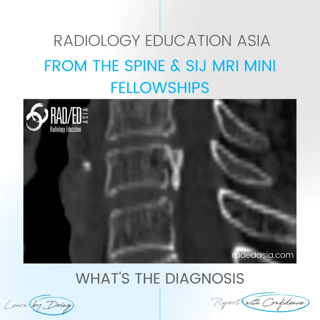
  • There is localised, linear ossification present at the posterior margin of the C6 vertebra.
  • This has a typical appearance of OPLL (Ossification of the Posterior Longitudinal Ligament).
  • The cervical spine is the most common location.
  • It is important to follow this up with an MRI of the cord to assess for cord compression and myelopathy.

Ossification present in the midline at the posterior body margin (Pink arrows).

If your Browser is blocking the video, Please view it on our YouTube Channel HERE

Learn more about SPINE Imaging in our ONLINE
Guided MRI Degenerative SPINE Mini-Fellowship.


More by clicking on the images below.

Axial CT image of the lumbar spine at the L4-L5 level showing focal calcification of the ligamentum flavum. Note both soft tissue thickening of the ligament flavum and the more dense calcification both contributing to right sided subarticular/lateral recess stenosis.
PAIN MANAGEMENT
arachnoiditis spine mri online radiology lumbar cord
RHEUMATOLOGY

#rheumatology #arthritis #rheumatologist #opll #ossification #degenerativedisease #orthopaedic #painphysician #chiropractic #chiropracter #physiotherapy #sportsmed #orthopaedic #mskmri #spondyloticmyelopathy #cordsignal #mricordradiology #radedasia #mri #spinemri #mskmri #mrispine #radiologyeducation #radiologycases #radiologist #radiologycme #radiologycpd #medicalimaging #imaging #radcme #spinedegeneration

error: Content is protected !!

Join our Newsletter

Stay tuned on new
Mini-Fellowships launches and learnings

Mini-Fellowships

Better Than Conferences

Join our WhatsApp Community

We help radiologists learn
by doing and report with confidence

This site is intended for Medical Professions only. Use of this site is governed by our Terms of Service and Privacy Statement which can be found by clicking on the links. Please accept before proceeding to the website.