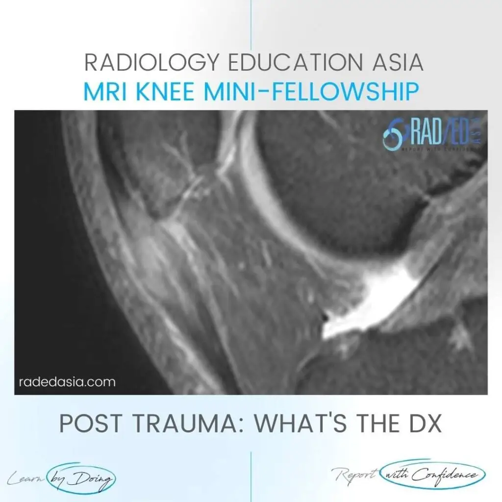
In this case we have Patellar tendon contusion and Patella contusion.
Most commonly this combined appearance would be of Patellar Tendonosis with reactive oedema in the patella and inflammatory change extending into Hoffa’s fat.
However the history of trauma in this patient is important in being able to differentiate the two.
.
WHAT ARE THE FINDINGS
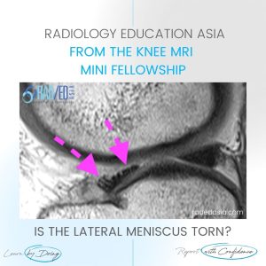
Increased PDFS signal and expansion proximal patellar tendon (Pink arrow) with,
Bone Marrow Oedema adjacent inferior pole patella (Blue arrow) and,
Oedema in the adjacent Hoffa’s Fat Pad.
PATELLAR TENDON CONTUSION ( MOVE SLIDER ICON TO LEFT)
Este sitio está destinado exclusivamente a las profesiones médicas. El uso de este sitio se rige por nuestras Condiciones de servicio y Declaración de privacidad, que puede consultar haciendo clic en los enlaces. Por favor, acéptelas antes de continuar en el sitio web.