ACROMIAL ACROMION SPUR MRI RADIOLOGY: HOW TO FIND ON CORONAL SCANS
ACROMION ACROMIAL SPUR MRI RADIOLOGY: WHAT TO LOOK FOR ON CORONAL SCANS
-
- What do Acromial spurs look like on MRI.
-
- Acromial / Acromion spurs can be small and difficult to see on MRI.
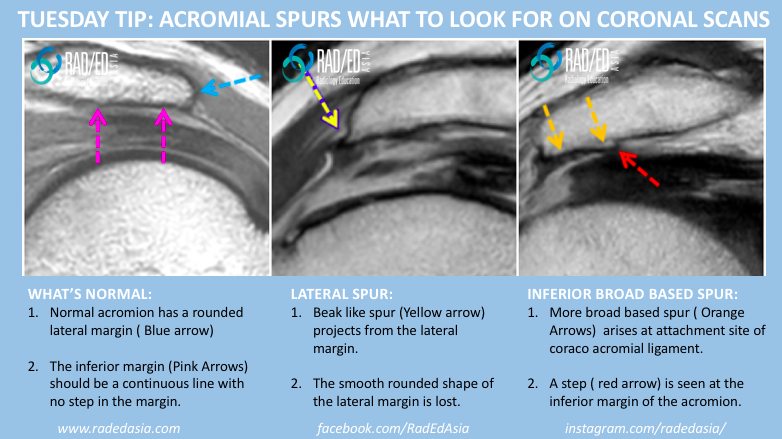
- Acromial spurs can be assessed on coronal and sagittal scans and this post looks at the MRI appearance of acromial spurs on coronal scans.


- Normal acromion has a rounded lateral margin (Blue arrow).
- The inferior margin (Pink Arrows) should be a continuous line with no step in the margin.

- Beak like spur (Yellow arrow) projects from the lateral margin.
- The smooth rounded shape of the lateral margin is lost.

- More broad based spur (Orange Arrows) arises at attachment site of coraco acromial ligament.
- A step (red arrow) is seen at the inferior margin of the acromion.

- Join our WhatsApp RadEdAsia community for regular educational posts at this link: https://bit.ly/radedasiacommunity
- Get our weekly email with all our educational posts: https://bit.ly/whathappendthisweek


