guillain-barre-syndrome-mri-findings-nerve-enhancement-spine-radiology-radedasia

MRI FINDINGS IN GUILLIAN BARRE SYNDROME

Guillain-Barré Syndrome (GBS) has distinct MRI findings in the spine. In this post we look at the characteristic MRI findings of GBS in the spine.

  • This is the most significant MRI finding in Guillain-Barré Syndrome.
     
  • Look for enhancement of the nerve roots in the cauda equina region on post-contrast T1-weighted images (Blue arrows axial and sagittal images).
     
  • Always fat saturate the post contrast T1 scans to make it easier to see the enhancement.

  • The enhancement signifies inflammation of the nerve roots, which is a characteristic feature of Guillain-Barré Syndrome.
     
  • This inflammation may cause the nerve roots to appear thicker than normal. However this is can be subjective and is not that common to see . In this case there is no appreciable swelling (Pink arrows).

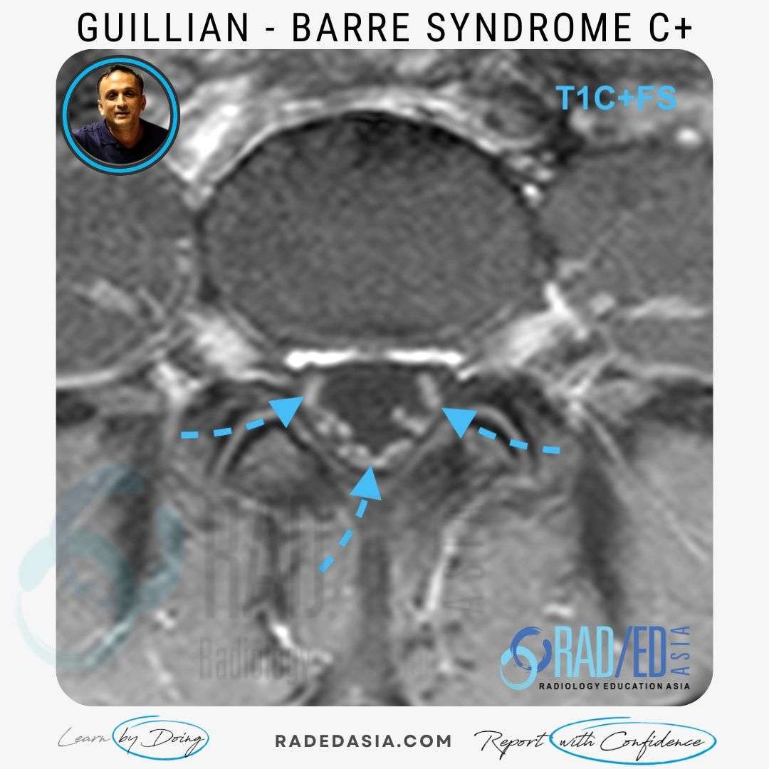
Typically, the enhancement of the nerve roots is symmetric (Blue arrows axial image), affecting nerve roots on both sides of the thecal sac.

  • GBS typically does not cause any other structural abnormalities in the spine.
     
  • If there are other abnormal findings in the spine consider other diagnoses.

  • Typically, the brain MRI is normal in Guillain-Barré Syndrome patients.
     
  • If there are other abnormal findings in the Brain, consider other diagnoses.

  • This is really up to the clinician and would only be done if the patient is not responding to treatment as expected.
     
  • In follow up scans look for a decrease in nerve root enhancement +/- reduction in nerve size if it was initially enlarged, indicating response to treatment.

No. Nerve root enhancement and thickening are non specific and can be seen in infection or malignancy amongst other causes. The clinical context of the scan is important in differentiating these. 

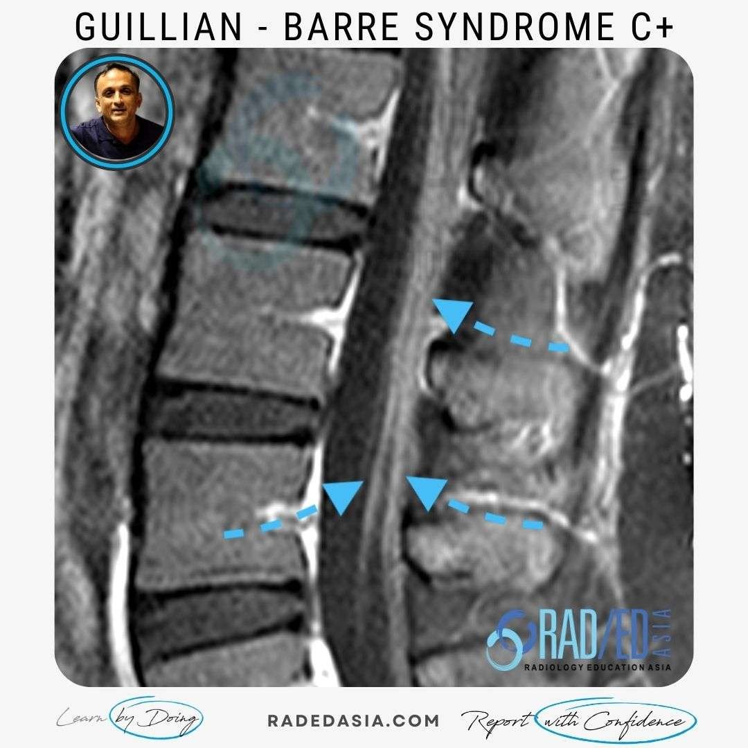
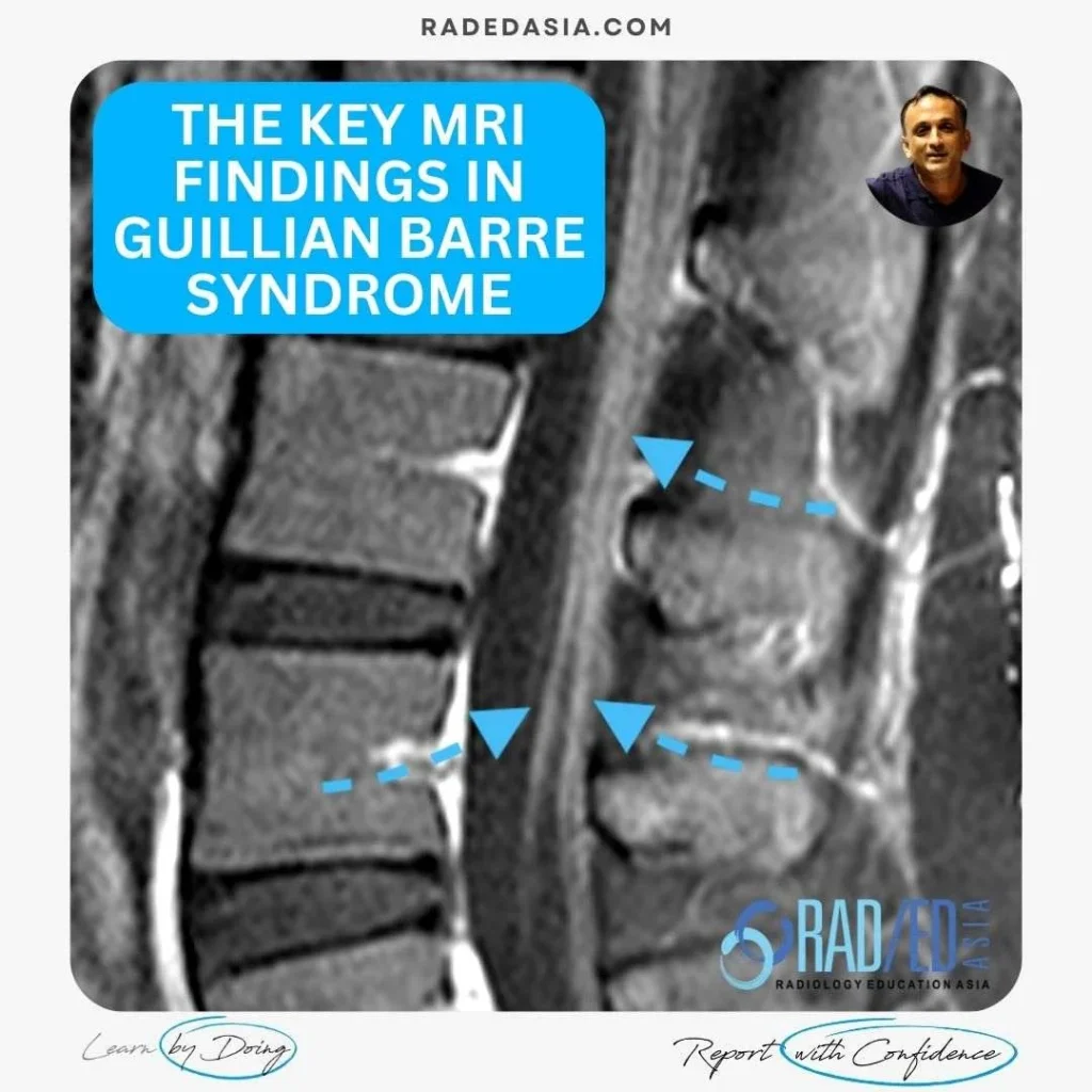
The most significant MRI finding in Guillain-Barré Syndrome is the enhancement of the nerve roots in the cauda equina region on post-contrast T1-weighted images.
.

The enhancement of the nerve roots can be visualized more easily by fat saturating the post-contrast T1 scans.

Yes, typically the enhancement of the nerve roots is symmetric, affecting nerve roots on both sides of the thecal sac.

Yes, typically the brain MRI is normal in Guillain-Barré Syndrome patients.

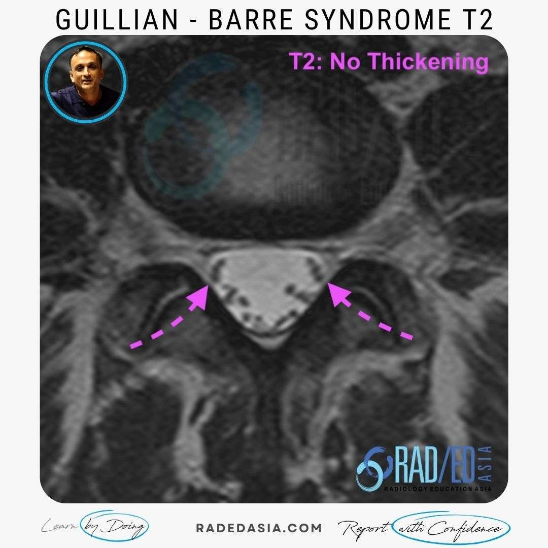
In follow-up MRI scans, a decrease in nerve root enhancement ( and size if the nerve roots were initially thickened)  should be looked for, indicating response to treatment.

Our CPD & Learning Partners

Learn more about this condition & How best to report it in more detail in our SPINE MRI Mini Fellowships.

Click on the image below for more information.

For all our other current MSK MRI & Spine MRI
Online Guided Mini Fellowships.


Click on the image below for more information.

#radedasia #mri #mskmri #radiology

This site is intended for Medical Professionals only. Use of this site is governed by our Terms of Service and Privacy Statement which can be found by clicking on the links. Please accept before proceeding to the website.
error: Right Click not available.