BASILAR INVAGINATION RADIOLOGY RHEUMATOID ARTHRITIS FIVE WAYS TO ASSESS
BASILAR INVAGINATION RADIOLOGY RHEUMATOID ARTHRITIS:
Subluxation at C1/2 in Rheumatoid Arthritis can occur in three directions.
- Axial (Horizontal).
- Vertical and,
- Lateral.
- Vertical subluxation is when C2 moves proximally towards the foramen magnum and is called Basilar Invagination or Basilar Impression.
- Basilar Invagination is when there is abnormal underlying bone and Basilar Impression is used when the underlying bone is normal.
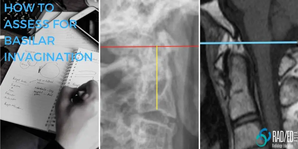
- Basilar invagination on X-ray, CT or MRI can be assessed in a number of ways the easiest being McRae's Line.
McRae's Line is drawn from the anterior to the posterior margin of the Occipital Foramen.
If the tip of the odontoid extends above this line, you can make a diagnosis of Basilar Invagination.
-
- Drawing McRae's Line is straight forward on CT and MRI but on a standard lateral X-ray its difficult to see the Occipital Foramen.
-
- So other methods (see below) have to be used to determine if the dens has migrated proximally.
-
- There are a number of other ways to assess for basilar invagination depending on whether you can adequately see the dens on the lateral x-ray or not.
- We know that drawing lines are the favorite activity of radiologists 🙂 but these are relatively straightforward and the images below can be used as a reference when required.
#radiology #radedasia #mri #spinemri #radiologyeducation #basilarinvagination #mcraeline #chamberlainline #mcgregorline #redlindjohnellmethod #clarkemethod #spondyloarthritis #ankylosingspondylitis #spinearthritis #radiologycases #radiologist #radiologycme #radiologycpd #medicalimaging #imaging #radcme #rheumatology #arthritis #rheumatologist #orthopaedic #painphysician #chiropractic #chiropracter #physiotherapy #sportsmed #orthopaedic #mskmri


