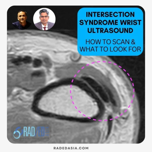
PAIN MANAGEMENT
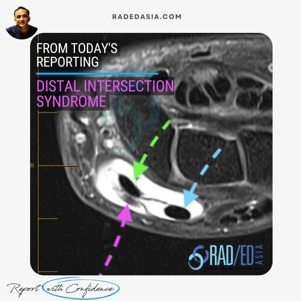
Distal intersection syndrome on MRI is relatively uncommon and occurs at the crossover of the extensor pollicis longus (EPL) over the extensor carpi radialis brevis (ECRB and ECRL) distal to Lister’s tubercle.
![]()
Lister’s tubercle acts as a pulley for the EPL tendon, which changes its angle of course, leading it to cross superficially over the second compartment tendons.
This can lead to increased friction between the tendons.
There is ALSO a potential communication foramen between the second and third compartments, which can allow inflammation to spread between them.
![]()
In this case there is significant tenosynovitis around the EPL (Pink arrow), ECRL (Green arrow) and ECRB (Blue arrow) with distension of the tendon sheaths of the second and third dorsal compartments.
![]()
There can also be additional findings of:
![]()
Radsource – MRI Features of Distal Intersection Syndrome.![]()
MRI typically shows tenosynovitis of the EPL and ECRL/ECRB, +/- peritendinous oedema, synovial thickening, and at times T2 hyperintensity at Lister’s tubercle.![]()
Proximal Intersection syndrome occurs 4-8 cm proximal to the wrist joint in the distal forearm and involves the first extensor compartment (abductor pollicis longus – APL and extensor pollicis brevis – EPB) crossing over the second compartment (ECRL and ECRB).
Distal intersection syndrome involves the second and third extensor compartments, whereas de Quervain’s tenosynovitis affects the first extensor compartment (abductor pollicis longus and extensor pollicis brevis).![]()
Fat-suppressed PD or T2-weighted and STIR sequences are ideal for identifying inflammation, peritendinous edema, and synovial thickening.
![]()


