- The medial patella plica is a developmental synovial membrane remnant.
- Can be seen in up to 30% of knees.
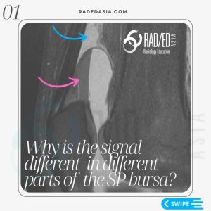
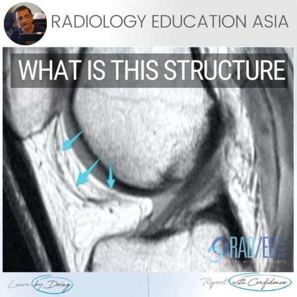
- It can be completely asymptomatic but can extend between the patella and trochlea and be compressed and cause pain. This is seen in so called Patella Plica Syndrome.
Â
- May also have chondromalacia in the adjacent patella and trochlea facet.
- A normal plica as shown is well defined and low signal.
Â
- It is found in the superomedial aspect of the joint and is best seen on axial scans.
Â
- With compression it will be thickened and ill defined and increase in signal.
Learn more about KNEE Imaging in our ONLINE or ONSITE
Guided MRI KNEE Mini-Fellowship.
More by clicking on the images below.
#radiology #radedasia #mri #kneemri #msk #mriknee #mskmri #radiologyeducation #radiologycases #radiologist #rads #radiologystudent #radiologycme #radiologycpd #medicalimaging #imaging #radcme #rheumatology #arthritis #rheumatologist #sportsmed #orthopaedic #physio #physiotherapist #kneeplica #plica #medialpatellaplica