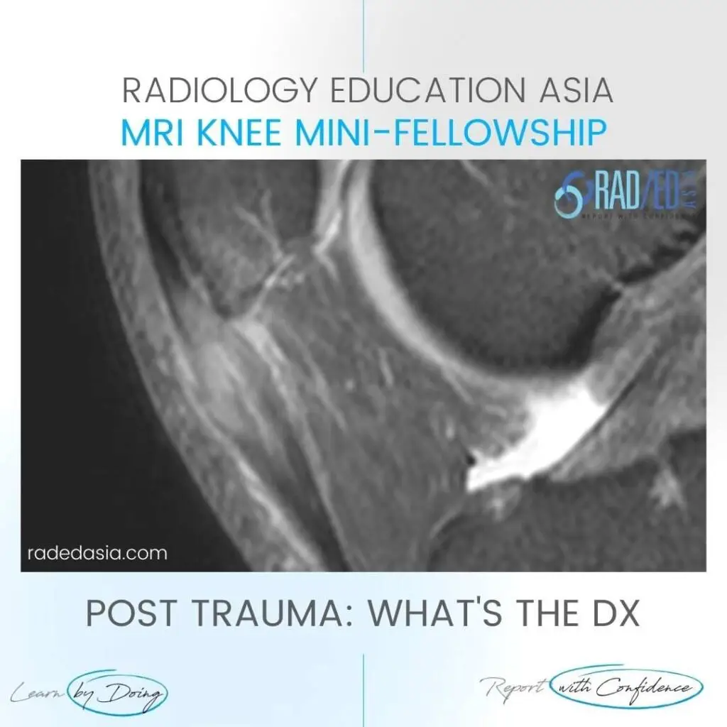
In this case we have Patellar tendon contusion and Patella contusion.
Most commonly this combined appearance would be of Patellar Tendonosis with reactive oedema in the patella and inflammatory change extending into Hoffa’s fat.
However the history of trauma in this patient is important in being able to differentiate the two.
.
WHAT ARE THE FINDINGS
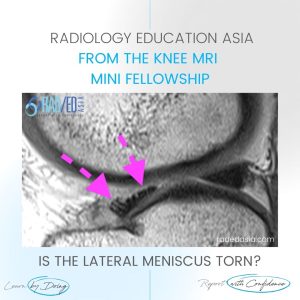
Increased PDFS signal and expansion proximal patellar tendon (Pink arrow) with,
Bone Marrow Oedema adjacent inferior pole patella (Blue arrow) and,
Oedema in the adjacent Hoffa’s Fat Pad.
PATELLAR TENDON CONTUSION ( MOVE SLIDER ICON TO LEFT)
This site is intended for Medical Professions only. Use of this site is governed by our Terms of Service and Privacy Statement which can be found by clicking on the links. Please accept before proceeding to the website.