The retrocalcaneal bursa lies anterior and deep to the Achilles tendon postero superior to the calcaneum.
The normal bursa has minimal fluid so on MRI you may see nothing or just a thin amount of fluid without synovial thickening.
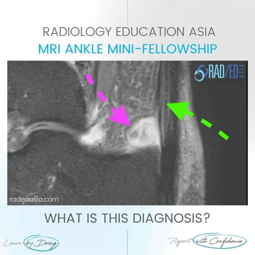
For retrocalcaneal bursitis on MRI look for contained fluid signal intensity in the expected location of the bursa.
In this case there is significant fluid in the bursa (Pink arrow) in keeping with bursitis.
The Achilles tendon is usually abnormal with tendonosis or tears. In this case, increased intermediate signal and expansion Achilles tendon (Green Arrow) in keeping with tendonosis.
This site is intended for Medical Professions only. Use of this site is governed by our Terms of Service and Privacy Statement which can be found by clicking on the links. Please accept before proceeding to the website.