
3 LATERAL KNEE FRACTURE: WHAT THEY ARE AND WHAT THEY MEAN
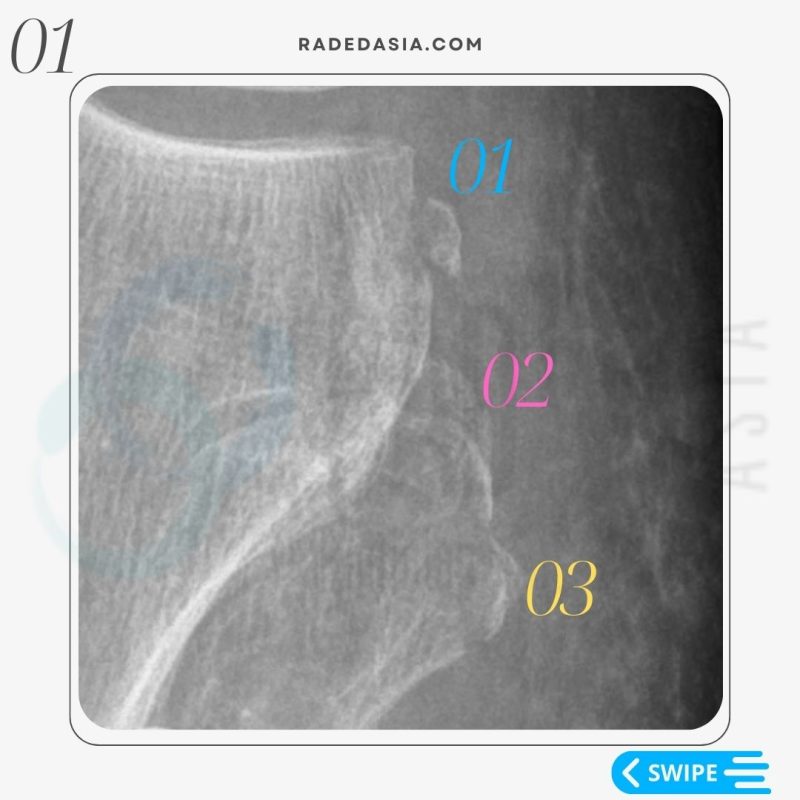
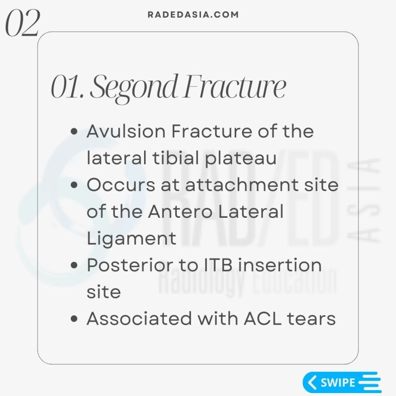
01: SEGOND FRACTURE
• Avulsion Fracture of the lateral tibial plateau.
• Occurs at attachment site of the Antero Lateral Ligament.
• Posterior to IT B insertion site.
• Associated with ACL tears.

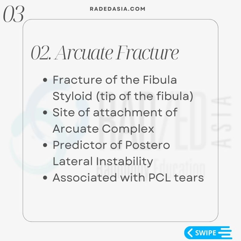
02: ARCUATE FRACTURE
• Fracture of the Fibula Styloid (tip of the fibula).
• Site of attachment of Arcuate Complex.
• Predictor of Postero-Lateral Instability.
• Associated with PCL tears.

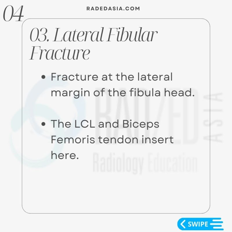
03: LATERAL FIBULAR FRACTURE
• Fracture at the lateral margin of the fibula head.
• The LCL and Biceps Femoris tendon insert here.

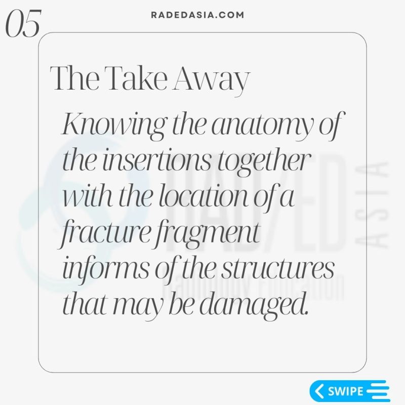
THE TAKEAWAY
Knowing the anatomy of the insertions together with the location of a fracture fragment informs of the structures that may be damaged.

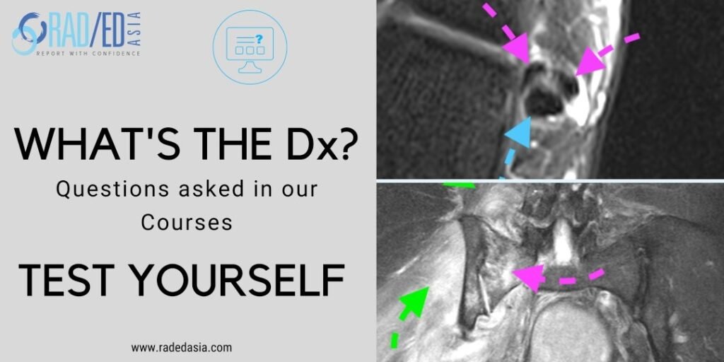
SUPRA PATELLAR PLICA: VIEW IMAGES
Learn more about this condition and how to best report it in more detail in our Guided KNEE MRI Mini Fellowship.
Click on the image below for more information.
- Join our WhatsApp RadEdAsia community for regular educational posts at this link: https://bit.ly/radedasiacommunity
- Get our weekly email with all our educational posts: https://bit.ly/whathappendthisweek


