radedasia

spine facet joint mri online radiology courses

SPINE MRI NERVE ROOT COMPRESSION ONLINE RADIOLOGY COURSE

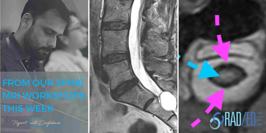
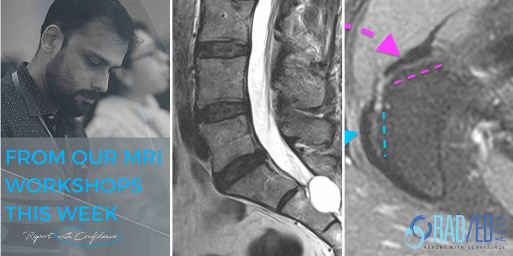
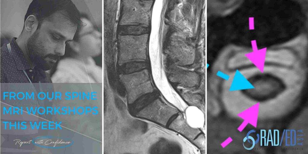
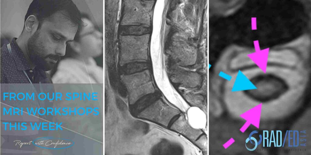
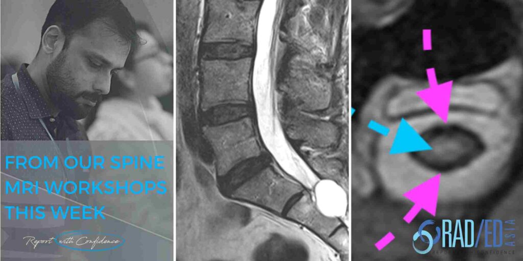
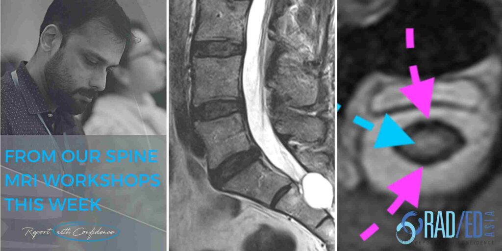
KEY POINTS FROM OUR SPINE MRI COURSES Spine MRI Nerve Root Compression Online Radiology Course Another three quick images with some basic key points from our online spine mri courses. Here we look at how to evaluate and describe a disc that extends into the foramen and its relationship to the exiting foraminal nerve looking […]

SPINE MRI NERVE ROOT COMPRESSION ONLINE RADIOLOGY COURSE Read More »

MRI LUMBAR FACET JOINT CYST RADIOLOGY: WHERE TO LOOK AND WHAT TO LOOK FOR (VIDEO)

LUMBAR SPINE FACET JOINT SYNOVIAL CYSTS MRI AND CT Lumbar Facet Joint Cysts MRI and CT: MRI Spine How to Assess for Facet Cysts and where to look. Today’s Tuesday Tip is a video on MRI of the Spine and Facet Cysts and in particular the MRI Spine Anatomy that relates to Spine Facet Cysts.

MRI LUMBAR FACET JOINT CYST RADIOLOGY: WHERE TO LOOK AND WHAT TO LOOK FOR (VIDEO) Read More »

learn radiology online msk mri shoulder ighl capsulitis radedasia

MRI ADHESIVE CAPSULITIS IGHL FROZEN SHOULDER (VIDEO)

Stay tuned on new Fellowships and learnings Subscribe what’s the dx? MRI ADHESIVE CAPSULITIS IGHL: MRI findings of Adhesive capsulitis also know as Frozen Shoulder In this post we look at the MRI Frozen Shoulder / MRI Adhesive Capsulitis. Frozen Shoulder or Adhesive capsulitis on MRI  is a common abnormality but is often missed because

MRI ADHESIVE CAPSULITIS IGHL FROZEN SHOULDER (VIDEO) Read More »

ct mri facet cysts mri spine how to mri facet cysts spine learn spine mri online radiology education asia

LUMBAR SPINE FACET JOINT SYNOVIAL CYST RADIOLOGY: MRI AND CT APPEARANCE (VIDEO)

LUMBAR SPINE FACET JOINT SYNOVIAL CYSTS MRI AND CT Today’s post is a video on MRI of the Spine and Facet Joints and in particular the MRI and CT appearance of Lumbar Spine Facet Joint Synovial Cysts. This approx 5 minute video below looks at What do spine facet joint cysts look like on MRI

LUMBAR SPINE FACET JOINT SYNOVIAL CYST RADIOLOGY: MRI AND CT APPEARANCE (VIDEO) Read More »

mri plantar fascia tear fasciitis plantar fascia anatomy heel pain mri mri online course radedasia

MRI PLANTAR FASCIA ANATOMY: WHAT ARE THE 3 BANDS OF THE PLANTAR FASCIA (VIDEO)

MRI PLANTAR FASCIA ANATOMY MRI PLANTAR FASCIA ANATOMY: NORMAL BANDS Today’s post is a video on MRI of the Plantar Fascia Anatomy. We are looking at how to identify the 3 bands of the Plantar Fascia and which band is most important in Plantar Fasciitis. If you find the video helpful, please subscribe to the

MRI PLANTAR FASCIA ANATOMY: WHAT ARE THE 3 BANDS OF THE PLANTAR FASCIA (VIDEO) Read More »

peroneal tubercle mri retrotrochlear eminence mri peroneal temdon tondonitis tear radiology education asia(1)

MSK MRI PERONEAL TUBERCLE & RETROTROCHLEAR EMINENCE MRI (VIDEO)

MRI PERONEAL TUBERCLE RETROTROCHLEAR EMINENCE MRI MSK How to identify the Peroneal tubercle and Retrotrochlear eminence on MRI: Today we are looking at MRI of the Peroneal Tubercle and the Retrotrochlear eminence. The importance of these structures is that when enlarged, a Peroneal Tubercle and less commonly the Retrotrochlear eminence, can result in Peroneal Tendonitis,

MSK MRI PERONEAL TUBERCLE & RETROTROCHLEAR EMINENCE MRI (VIDEO) Read More »

spine mri msk online radiology courses radedasia

LUMBAR SPINE ENHANCEMENT MRI ONLINE RADIOLOGY COURSES: KEY POINTS FROM OUR SPINE MRI COURSES

KEY POINTS FROM OUR SPINE MRI COURSES Spine MRI Online Radiology Course: Normal Spinal Enhancement Another three quick images with some basic key points from our online spine MRI courses. Here we look at areas of normal enhancement that can be mistaken for pathology: Normal enhancement of the anterior epidural plexus which can be mistaken

LUMBAR SPINE ENHANCEMENT MRI ONLINE RADIOLOGY COURSES: KEY POINTS FROM OUR SPINE MRI COURSES Read More »

ankylosing spondylitis sacroileitis sacro iliac joint ct mri online spine radiology courses

ANKYLOSING SPONDYLITIS SACROILIITIS CT CHANGES SACRO ILIAC JOINT SPINE MRI RADIOLOGY COURSE

ANKYLOSING SPONDYLITIS SACROILIITIS CT CHANGES MRI Ankylosing Spondylitis Sacroiliitis CT changes MRI Online Radiology Course Another three quick images with some basic key points from our online spine MRI courses. Here we look at the changes seen on CT at the Sacro Iliac Joint. CT ANKYLOSING SPONDYLITIS SACRO ILIAC JOINT SCLEROSIS AND EROSIONS Bony erosions

ANKYLOSING SPONDYLITIS SACROILIITIS CT CHANGES SACRO ILIAC JOINT SPINE MRI RADIOLOGY COURSE Read More »

schmorl nodes spine mri online radiology courses

SCHMORL\’S NODES SPINE MRI ONLINE RADIOLOGY COURSE LEARN KEY POINTS

KEY POINTS FROM OUR SPINE MRI COURSES Schmorl’s Node Spine MRI Online Radiology Course Acute and Chronic Schmorl’s nodes. Another three quick images with some basic key points from our online spine MRI courses. ACUTE SCHMORL'S NODE MRI Acute schmorl’s nodes demonstrate oedema at their margins. This is usually limited to around the circumference of

SCHMORL\’S NODES SPINE MRI ONLINE RADIOLOGY COURSE LEARN KEY POINTS Read More »

arachnoiditis spine mri online radiology courses radedasia

ARACHNOIDITIS MRI SPINE ONLINE RADIOLOGY COURSE

KEY POINTS FROM OUR SPINE MRI COURSES Arachnoiditis Spine MRI Online Radiology Course What are some of the common appearance of arachnoiditis on MRI? Another three quick images with some basic key points from our online spine MRI courses. ARACHNOIDITIS MRI ASYMMETRIC DISTRIBUTION WHAT'S THE SIGNIFICANCE OF ASYMMETRY? The distribution of nerve roots in the

ARACHNOIDITIS MRI SPINE ONLINE RADIOLOGY COURSE Read More »

error: Content is protected !!

Join our Newsletter

Stay tuned on new
Mini-Fellowships launches and learnings

Mini-Fellowships

Better Than Conferences

Join our WhatsApp Community

We help radiologists learn
by doing and report with confidence

This site is intended for Medical Professions only. Use of this site is governed by our Terms of Service and Privacy Statement which can be found by clicking on the links. Please accept before proceeding to the website.