MENISCUS TEAR MRI KNEE: MISSING MENISCI WHAT TO LOOK FOR
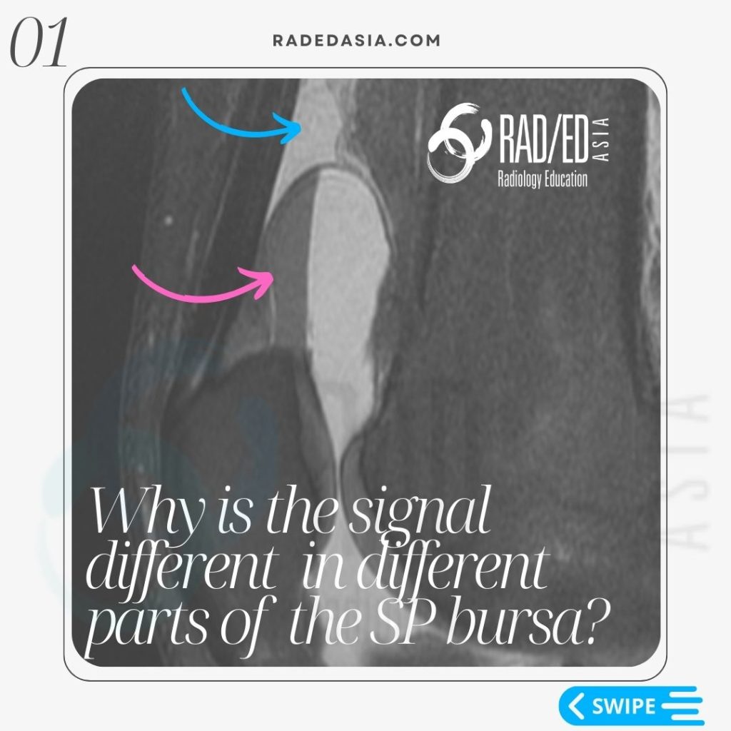
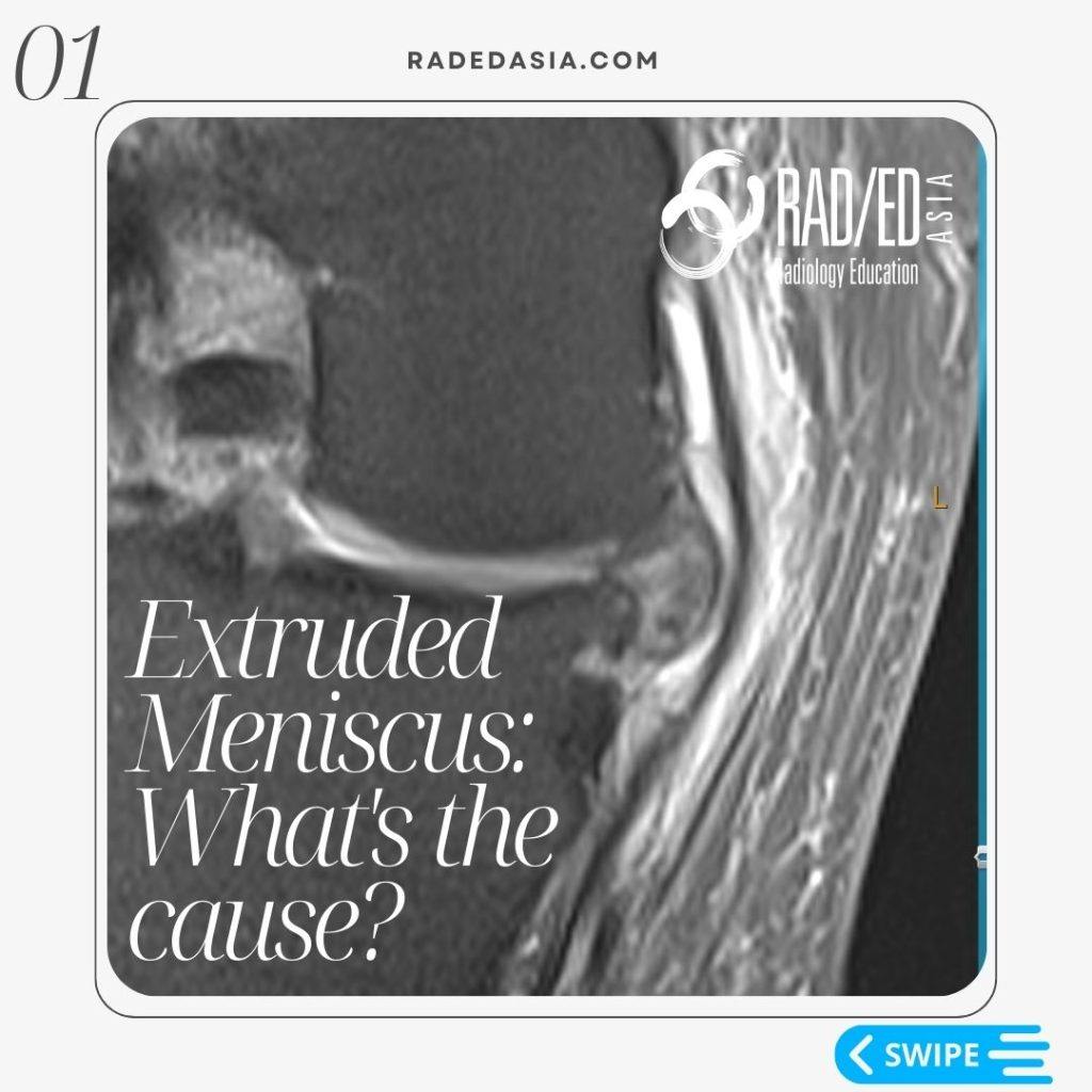
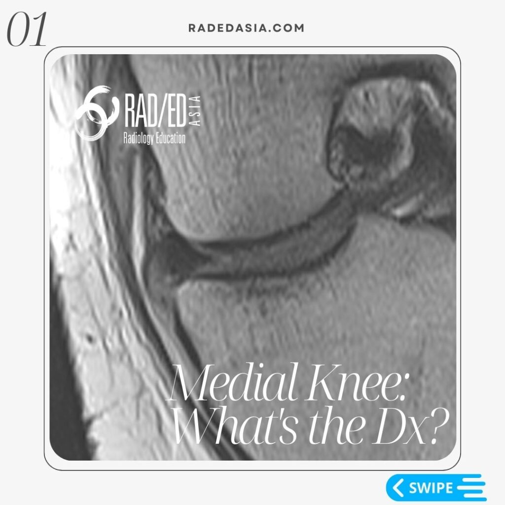
Stay tuned on new Fellowships and learnings Subscribe The Dx / Knee MENISCUS TEAR MRI KNEE WHAT TO LOOK FOR In a Normal meniscus the superior and inferior halves should be symmetric. In all three cases there is localised loss of ” volume” of the meniscus. This gives the meniscus an asymmetric look. Asymmetry in […]
MENISCUS TEAR MRI KNEE: MISSING MENISCI WHAT TO LOOK FOR Read More »