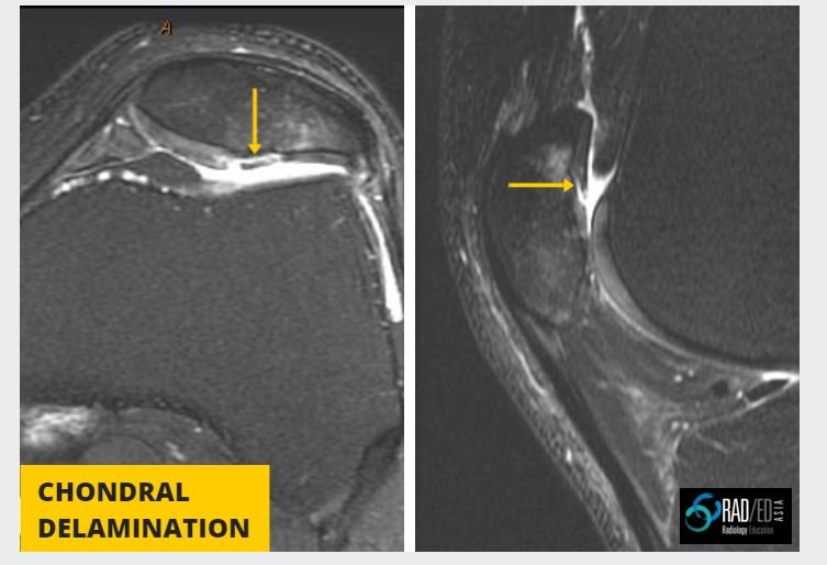
CHONDRAL DELAMINATION CARTILAGE MRI: Quick Review
CHONDRAL DELAMINATION CARTILAGE MRI
Chondral Delamination on MRI is not common but is seen often enough that we need to be aware of it and What to look for on MRI.

What is it? Chondral delamination is when cartilage separates and lifts off from its attachment with the cortex.
Why is it important? It can progress to complete loss of cartilage in that region.
Location: Most commonly seen in knee and ankle. Other joints uncommon.


- In chondral delamination, cartilage is elevated from the underlying subchondral bone.
- Look for a rim of high signal T2 (which is fluid) between subchondral bone and cartilage.
- The cartilage delamination can be associated with an adjacent area of full thickness cartilage loss or can be isolated and surrounded by normal cartilage.

Once delamination occurs it can progressively undermine adjacent cartilage until eventually cartilage falls off and we have areas of full thickness cartilage loss.
 Image above Progressive chondral delamination with eventual full thickness cartilage loss:
1. Commencing in July 2013 with a full thickness chondral fissure and early delamination to,
2. May 2015 Widening of area of chondral fissure and increasing delamination to,
3. Jan 2016 where delamination has converted to larger area of full thickness cartilage loss.


