mri-cartilage-early-changes-delamination-radedaia-radiology-education-asia

MRI CARTILAGE CHONDRAL DAMAGE: CARTILAGE DELAMINATION & EARLY CARTILAGE CHANGES

MRI CARTILAGE CHONDRAL DAMAGE & LESIONS: What to look for and How to report.


What are the early changes of chondral damage we should see and report. This post covers the 3 Things to look for.


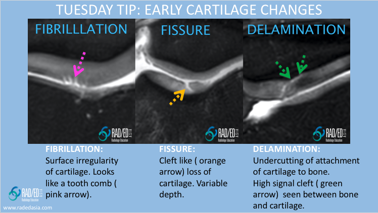
MRI CARTILAGE CHONDRAL DAMAGE: 3 THINGS TO LOOK FOR

mri cartilage delamination radiology education asia

Surface irregularity of cartilage. Looks like a tooth comb (Pink arrow).

Cleft like (Orange arrow) loss of cartilage. Variable depth.

    • Undercutting of attachment of cartilage to bone.


  • High signal cleft (Green arrow) seen between bone and cartilage.

#radiology #radedasia #mri #kneemri #msk #mriknee #mskmri #kneecartilage #cartilagedelamination #chondral #radiologyeducation #radiologycases #radiologist #rads #radiologystudent #radiologycme #radiologycpd #medicalimaging #imaging #radcme #rheumatology #arthritis #rheumatologist #sportsmed #orthopaedic #physio #physiotherapist 

#radedasia #mri #mskmri #radiology

This site is intended for Medical Professionals only. Use of this site is governed by our Terms of Service and Privacy Statement which can be found by clicking on the links. Please accept before proceeding to the website.
error: Right Click not available.