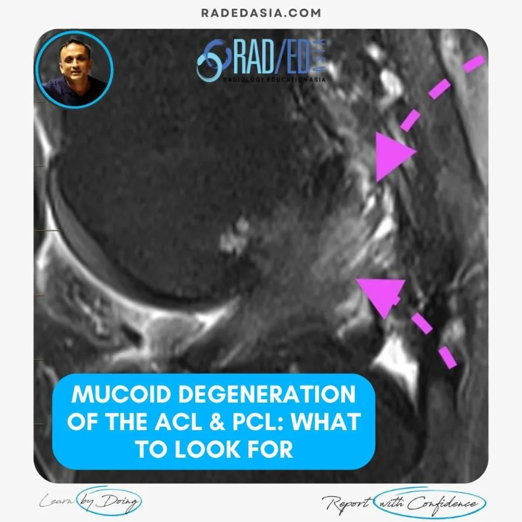
ACL & PCL MUCOID DEGENERATION MRI FINDINGS
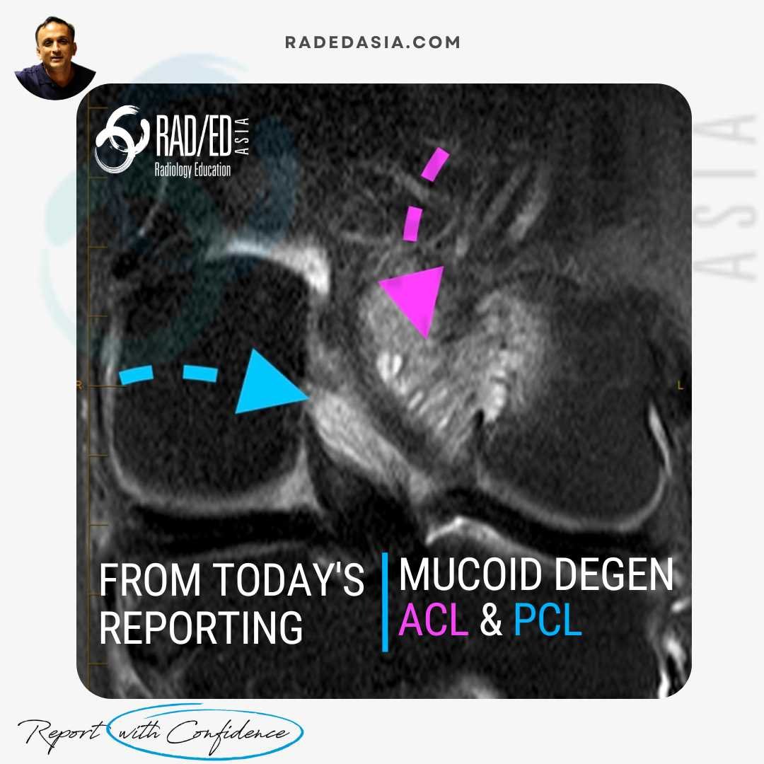
Mucoid degeneration can affect both the anterior cruciate ligament (ACL) and posterior cruciate ligament (PCL). Mucoid degeneration is often confused with ACL tears and this post looks at the main MRI features of ACL and PCL mucoid degeneration that will help to differentiate from tears.
WHAT ARE THE FINDINGS?
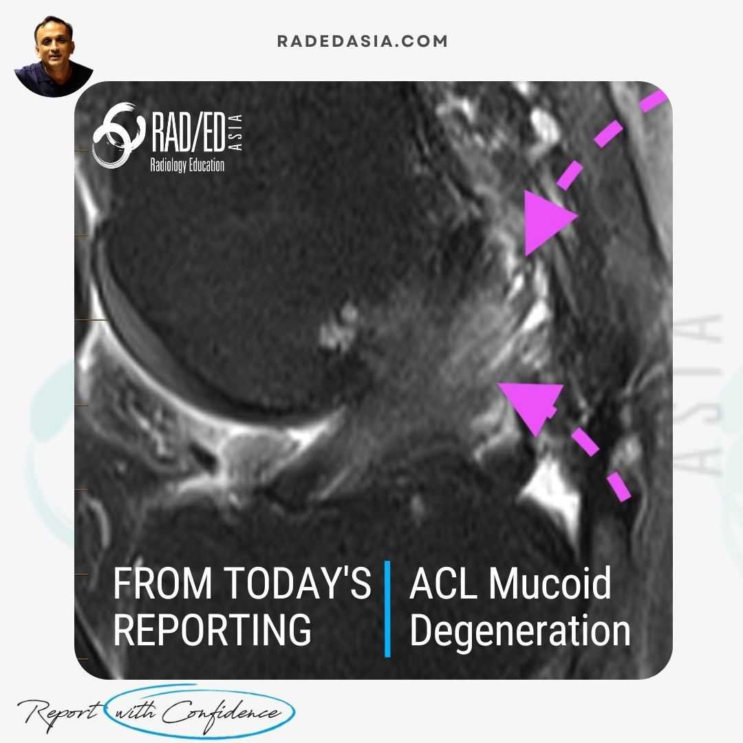
- The ACL is enlarged and hyperintense but there is continuity of fibres and normal alignment.
- The enlargement is more prominent proximally giving it the appearance of a celery stalk (celery stalk sign of ACL mucoid degeneration).
SUMMARY:
This is a characteristic appearance of ACL mucoid degeneration. Look for,
- Expanded hyperintense ACL.
- Fibres are continuous.
- Alignment of fibres is normal.

- Ligament thickening.
- "Celery stalk" sign.
- Increased T2 Signal intensity.
- Preserved fibres which are in continuity.
- Normal alignment of the ACL or PCL.
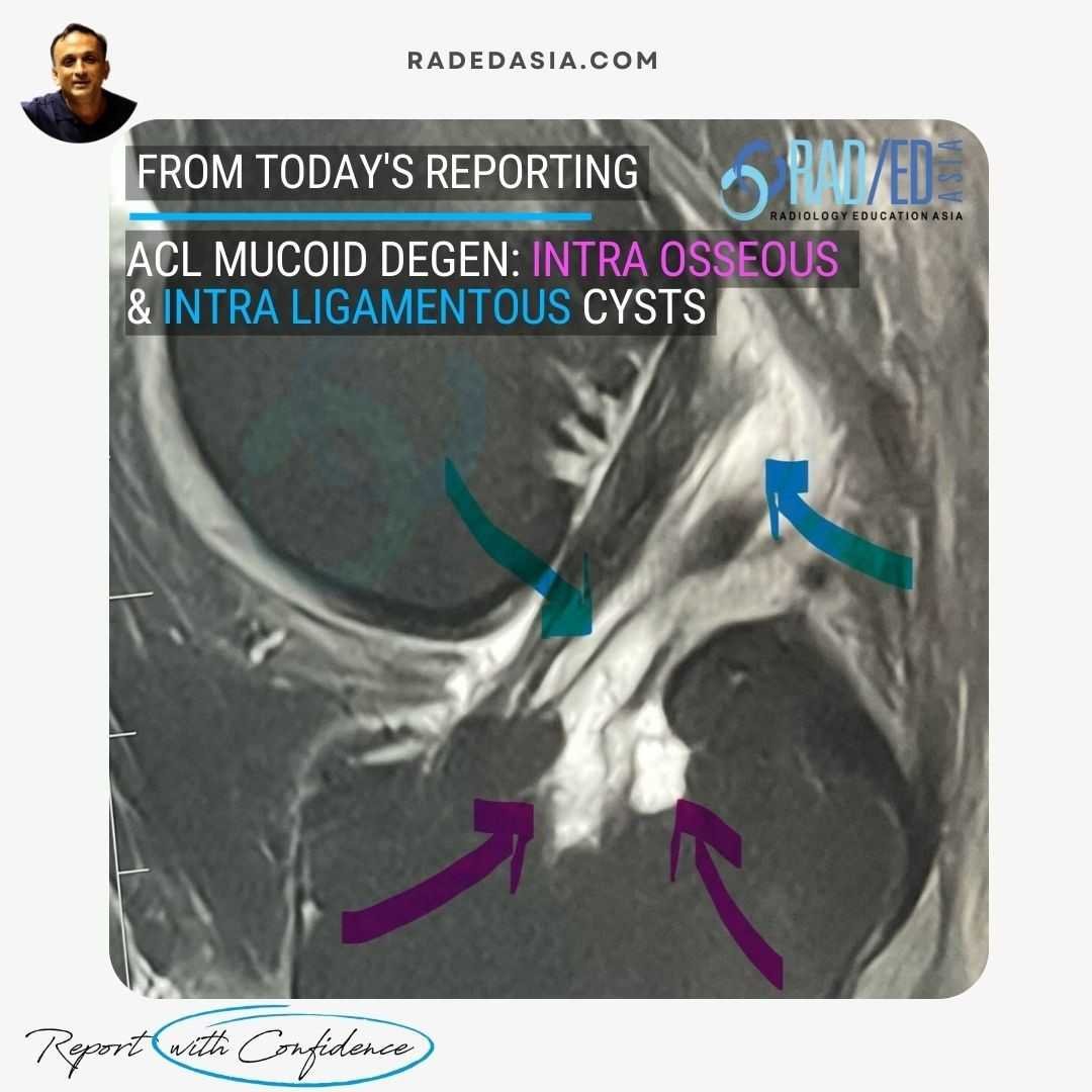
- Intra ligamentous cysts of variable size.
- Intra osseous extension of the cysts.

Learn more about this condition & How best to report it in more detail in our Online Guided KNEE MRI Mini Fellowship.
More by clicking on the images below.
For all our other current MSK MRI & Spine MRI
Online Guided Mini Fellowships.
Click on the image below for more information.
- Join our WhatsApp RadEdAsia community for regular educational posts at this link: https://bit.ly/radedasiacommunity
- Get our weekly email with all our educational posts: https://bit.ly/whathappendthisweek


