![Axial CT image of the lumbar spine at the L4-L5 level showing focal calcification of the ligamentum flavum. Note both soft tissue thickening of the ligament flavum and the more dense calcification both contributing to right sided subarticular/lateral recess stenosis.](https://radedasia.com/wp-content/uploads/2025/02/ct-imaging-ligamentum-flavum-hypertrophy-thickening-calcification-radedasia-1024x1024.webp)
- 8th March 2025
SGD$695.00
CT OF LIGAMENTUM FLAVUM THICKENING/HYPERTROPHY AND CALCIFICATION/OSSIFICATION
![](https://radedasia.com/wp-content/uploads/2024/09/REA-Logo-Transparent-BG.png)
FROM TODAY'S REPORTING
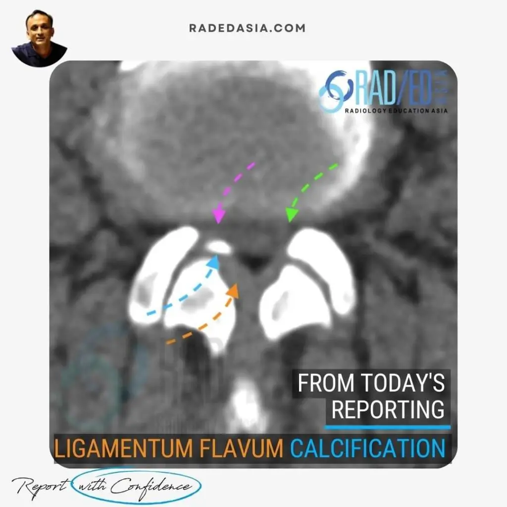
LIGAMENTUM FLAVUM CALCIFICATION
Â
Combination of thickened ligamentum flavum (Orange arrow) and Localized calcification of the flavum (Blue arrow) results in right sided Subarticular/ lateral recess stenosis (Pink arrow). Compare recess with left side (Green arrow) which is not narrowed. Canal stenosis also present.
Â
We look at all of these topics in more detail in our Guided SPINE & SIJ Imaging Mini Fellowships.
Click on the image below for more information.
- Join our WhatsApp RadEdAsia community for regular educational posts at this link: https://bit.ly/radedasiacommunity
- Get our weekly email with all our educational posts: https://bit.ly/whathappendthisweek
- 8th March 2025
SGD$695.00