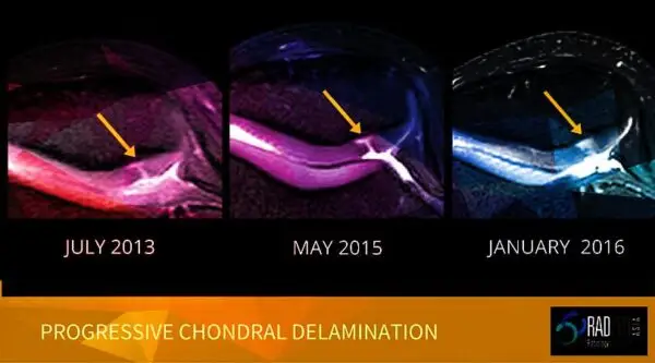
CARTILAGE
Shoulder cartilage MRI interpretation of early changes can be difficult because of the thinness of the cartilage, making the detection of early changes, such as cartilage fissures and delamination, less commonly seen than in the knee.
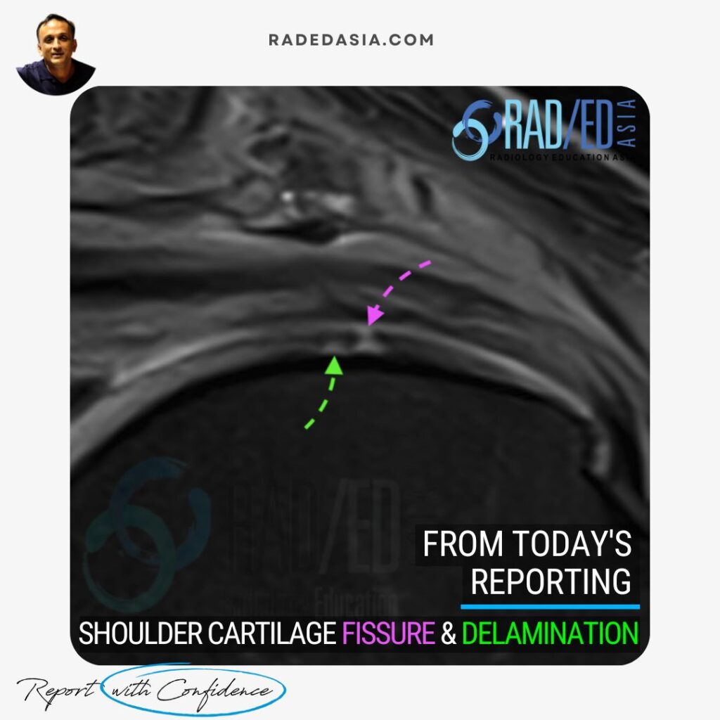
This case demonstrates a localized area of full-thickness fissuring (Pink arrow), accompanied by cartilage delamination (Green arrow). Notably, a high signal tract is observed deep to the cartilage, delineating the separation between the cartilage and bone.
Fluid traversing cartilage in a fissure or at the delamination interface is best seen on proton density fat-saturated (PDFS) and T2 fat-saturated (T2FS) sequences.![]()
Cartilage fissuring and delamination is commonly seen in the knee but its visualization in the shoulder is relatively uncommon due to the thin shoulder cartilage. Main finding to look for is high fluid signal traversing through cartilage for fissuring or running as an interface between cartilage and bone for delamination.![]()
Article: “Imaging of Glenohumeral Cartilage: Normal Anatomy and Pathological Findings” from Radiographics Journal, Read HERE![]()
The primary challenge is the thinness of the cartilage, making it difficult to visualize subtle changes like fissures and early delamination compared to joints like the knee.![]()
Delamination appears as a high signal tract on fluid-sensitive sequences (PDFS/T2FS) between the cartilage and bone.
![]()
Proton density fat-saturated (PDFS) and T2 fat-saturated (T2FS) sequences are optimal for visualizing fluid traversing cartilage defects and highlighting delamination.
![]()