
All Spanish Blogs
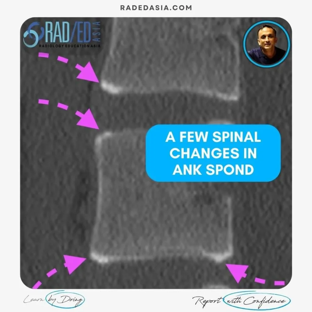
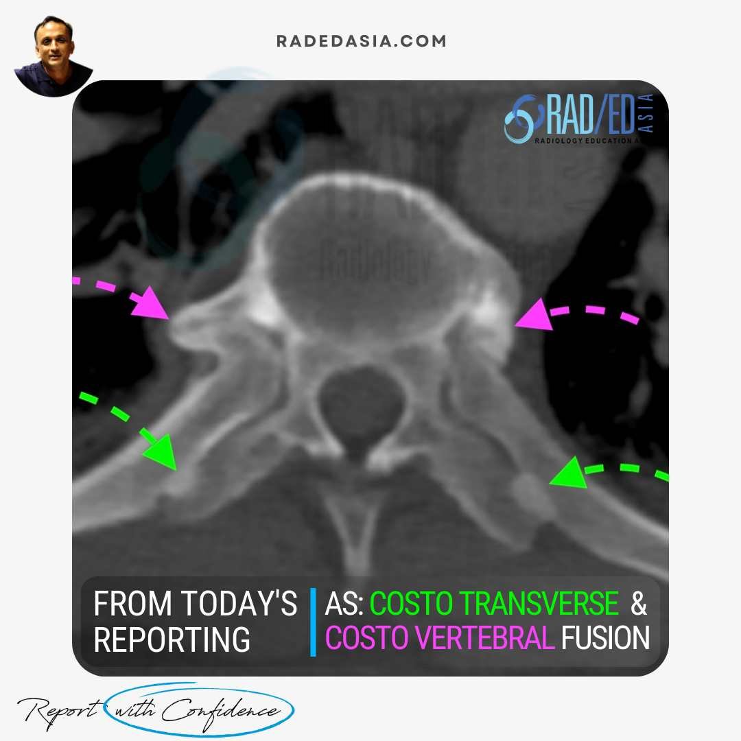
Ankylosing spondylitis (AS), in the spine can involve the anterior and posterior elements. It usually commences anteriorly and as the disease progresses, posterior changes are seen. In these images we look at:
![]()
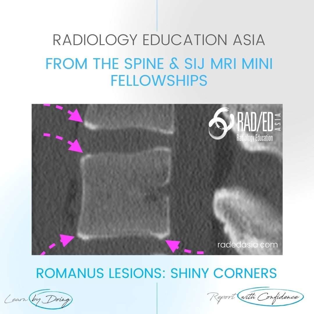
WHAT ARE THE FINDINGS?
The CT demonstrates sclerosis and some rounding of the corners of the vertebral bodies in keeping with Romanus Lesions in Ankylosing Spondylitis. Shiny corners refer to the sclerosis seen on CT.
![]()
CT Spine changes in Ankylosing Spondylitis:
Some early changes involving the vertebral body in ankylosing spondylitis:
![]()
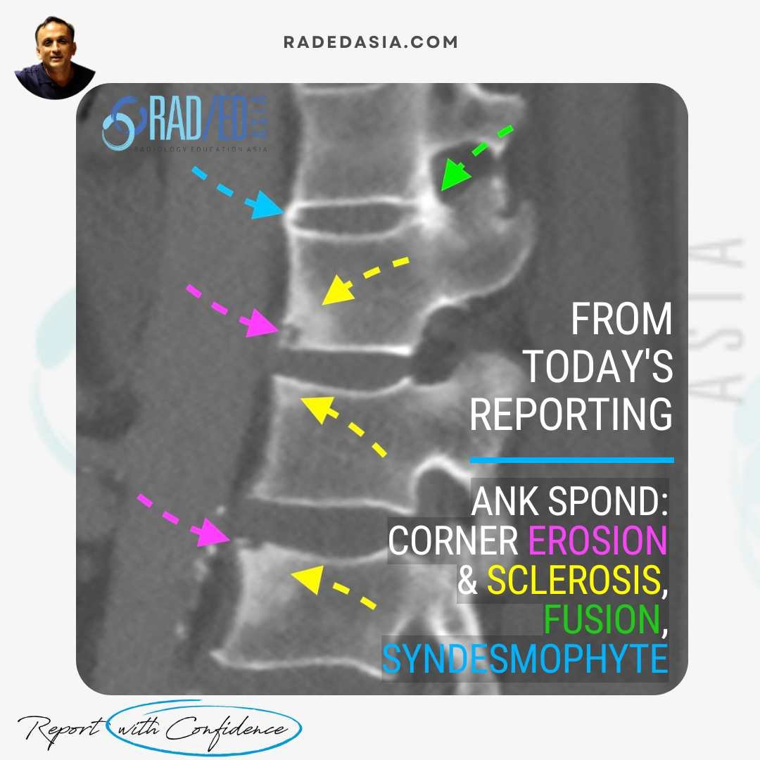
WHAT ARE THE FINDINGS?
![]()
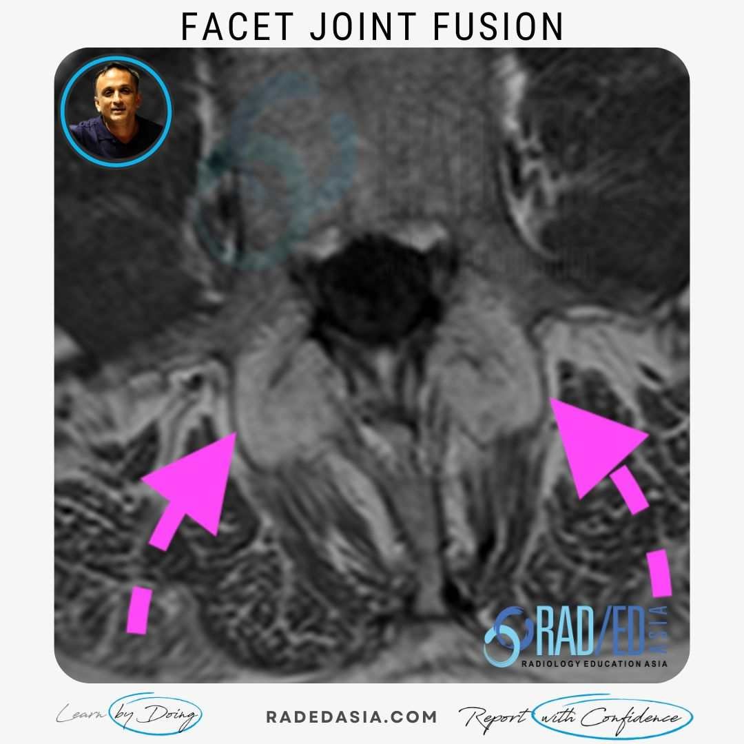
WHAT ARE THE FINDINGS?
![]()


