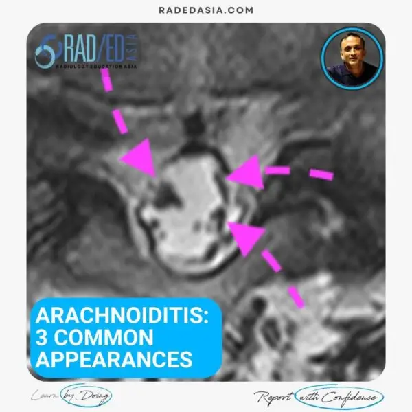

ARACHNOIDITIS PATTERNS ON MRI
Arachnoiditis in the lumbar spine on MRI is not uncommon to see and is easy to diagnose if you know what to look for. In this post we look at What is Normal, why does Arachnoiditis occur and What to look for.
![]()
- Arachnoiditis is not uncommon to see in the lumbar spine. But the most important thing about being able to recognize arachnoiditis is to first know the normal distribution of nerve roots in the spinal canal.
- The normal distribution is a bit like the horse's tail in that more proximally the nerve roots are together and as you go more distally, they separate.
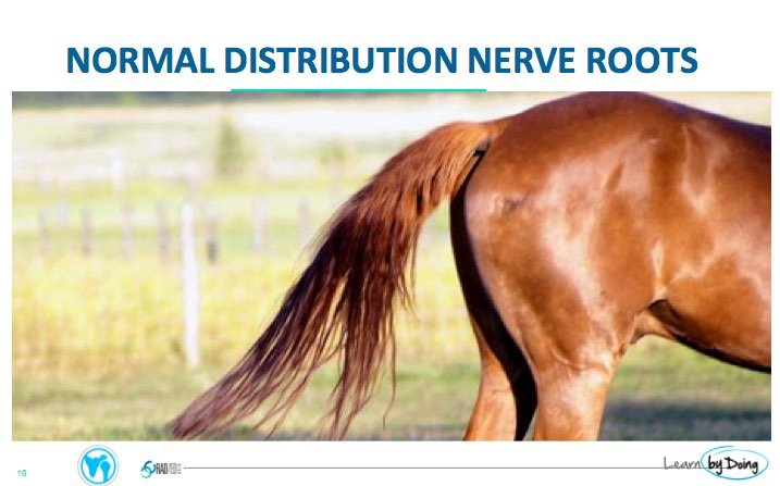

Image Above: The position of the nerve roots depends on what level you are looking at. However, it must be symmetrical on both sides whatever level you are looking at.
![]()
MECHANISM OF ARACHNOIDITIS:
Arachnoiditis commonly is due to Acute inflammation/infection. Hemorrhagic and chemical arachnoidits can also occur but are less common.
There is a sequence of pathological changes .
• Exudate and fibrin deposited on nerve sheaths and arachnoid.
• Collagen forms.
• Nerve roots stick together.
• Subarachnoid space becomes septated and cystic.

![]()
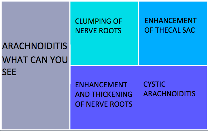
ARACHNOIDITIS PATTERNS:
There are a number of patterns of arachnoiditis. They can occur on their own or as a combination.
- Clumping of Nerve roots.
- Enhancement of the Thecal Sac.
- Enhancement & thickening of Nerve roots.
- Cystic Arachnoiditis.
![]()
- Clumping of nerve roots is due to a few nerve roots becoming attached to each other. There are multiple patters to this (like below) as it depends on which nerve roots attach to each other.
- The main appearance to look for is asymmetry of the distribution of nerve roots.

![]()
Thickening of individual nerve roots is due to inflammatory infiltrate lining the nerve roots. A similar appearance will also be seen with malignant arachnoiditis.

![]()
A blocked thecal sac in arachnoiditis is due to the nerve roots all clumped and attached together. This results in a solid appearing structure that fills the thecal sac.

![]()
Nerve roots can enhance in acute and subacute arachnoiditis. However the enhancement is not specific as it can be seen in inflammatory, infective or malignant causes. 
![]()
The thecal sac can be affected in two ways.
- Nerve roots stuck to the margins of the sac resulting in a so called empty thecal sac.
- Cystic changes in the thecal sac resulting in Cystic Arachnoiditis.

Image above:
- Left image demonstrates the normal distribution of nerve roots in the lower lumbar spine.
- The central image demonstrates nerve roots peripherally distributed against the thecal sac margins.
- Right image demonstrates an empty sac where it looks like there are no nerve roots. The nerve roots are present but are flattened against the walls of the sac and are difficult to separate on imaging.
![]()
Arachnoiditis can result in septations in the thecal sac resulting in intra dural arachnoid cyst formation.
Look for cystic type changes in the thecal sac with thickened walls.

Cytsic arachnoiditis can also result in cord oedema . See more on this by clicking on the image below.

![]()