Chris Ong

annular fissure tear mri radiology lumbar spine radedasia

ANNULAR FISSURE TEAR MRI RADIOLOGY LUMBAR SPINE (VIDEO)

Stay tuned on new Fellowships and learnings Subscribe The Dx / Spine Degeneration ANNULAR FISSURE TEAR MRI RADIOLOGY HOW DO YOU FIND AN ANNULAR FISSURE ANNULAR FISSURE ON MRI Annular fissures on MRI are relatively common. Whilst they don’t result in radiculopathy, they can be a cause of back pain and are important to diagnose […]

ANNULAR FISSURE TEAR MRI RADIOLOGY LUMBAR SPINE (VIDEO) Read More »

ankle impingement radiology anterior xray mri radedasia

ANKLE IMPINGEMENT RADIOLOGY ANTERIOR X-RAY (VIDEO)

Stay tuned on new Fellowships and learnings Subscribe The Dx / Ankle ANKLE IMPINGEMENT RADIOLOGY ANTERIOR X-RAY DISCUSSION Ankle impingement can occur at multiple sites. One of the more common ones is Anterior. The presence of Ankle impingement is ultimately a clinical diagnosis which is based on the patient’s symptoms and imaging findings, not just

ANKLE IMPINGEMENT RADIOLOGY ANTERIOR X-RAY (VIDEO) Read More »

scapula normal variant keats shoulder mri

NORMAL SCAPULA VARIANT SHOULDER MRI RADIOLOGY (VIDEO)

Stay tuned on new Fellowships and learnings Subscribe The Dx / Shoulder NORMAL SCAPULA VARIANT: CLASP LIKE SUPERIOR MARGIN FINDINGS Well corticated bridge of bone (Pink arrows) that lies on the superior margin of the scapula with a bone defect inferior to it. DISCUSSION This is a normal variant. Its described in Keats as a

NORMAL SCAPULA VARIANT SHOULDER MRI RADIOLOGY (VIDEO) Read More »

spine trauma radiology facet dislocation subluxation perched locked ct imaging radedasia

SPINE TRAUMA RADIOLOGY: HOW TO Dx A FACET DISLOCATION ON CT (VIDEO)

Stay tuned on new Fellowships and learnings Subscribe The Dx / Spine SPINE TRAUMA RADIOLOGY: FACET DISLOCATION ON CT FACET DISLOCATION ON CT: Spinal trauma can result in a number of abnormalities of the facet joints ranging from Diastasis, Subluxation, Perched Facets and eventually to Dislocation or Locked Facets. In this short video we look

SPINE TRAUMA RADIOLOGY: HOW TO Dx A FACET DISLOCATION ON CT (VIDEO) Read More »

myelomalacia-spondylotic-myelopathy-radiology-education-asia

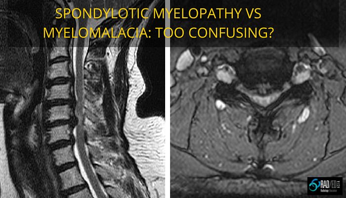
SPONDYLOTIC MYELOPATHY vs MYELOMALACIA: NO MORE CONFUSION

Stay tuned on new Fellowships and learnings Subscribe The Dx / Spine MYELOMALACIA vs SPONDYLOTIC MYELOPATHY Spondylolytic myelopathy and Myelomalacia Spondylolytic myelopathy and Myelomalacia are quite different but there is often confusion between the use of the terms. Let’s look at what they are and how to differentiate them. SPONDOLYTIC MYELOPATHY Refers to increased T2

SPONDYLOTIC MYELOPATHY vs MYELOMALACIA: NO MORE CONFUSION Read More »

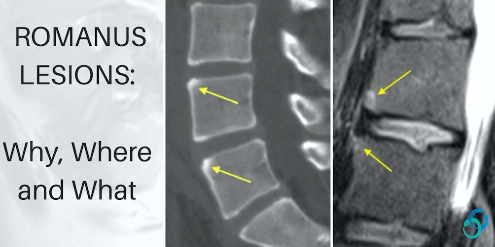
THE ROMANUS LESION: WHY, WHERE AND WHAT

Stay tuned on new Fellowships and learnings Subscribe The Dx / Spine ROMANUS LESION CORNER INFLAMMATORY LESION MRI CT The Romanus lesion (which is now called a Corner Inflammatory Lesion) is one of the earliest signs in the spine of inflammatory spondyloarthritis. ROMANUS LESION / CORNER INFLAMMATORY LESIONS The Romanus lesion represents inflammatory changes at

THE ROMANUS LESION: WHY, WHERE AND WHAT Read More »

error: Content is protected !!

Join our Newsletter

Stay tuned on new
Mini-Fellowships launches and learnings

Mini-Fellowships

Better Than Conferences

Join our WhatsApp Community

We help radiologists learn
by doing and report with confidence

This site is intended for Medical Professions only. Use of this site is governed by our Terms of Service and Privacy Statement which can be found by clicking on the links. Please accept before proceeding to the website.